The use of artificial intelligence to identify ophthalmic biomarkers in cardiovascular disease and stroke: a narrative review
Diogo Gonçalves dos Santos Martins, Thiago Goncalves dos Santos Martins, Paulo Schor

TL;DR
This paper reviews how artificial intelligence can detect eye-related signs of heart disease and stroke, offering non-invasive diagnostic insights.
Contribution
The paper systematically reviews AI applications in identifying ophthalmic biomarkers for cardiovascular disease and stroke.
Findings
AI effectively identifies non-invasive biomarkers from retinal exams for cardiovascular disease and stroke.
A total of 14 studies were critically analyzed for AI's role in ophthalmic biomarker detection.
Findings enhance understanding of the pathophysiology of cardiovascular disease and stroke.
Abstract
Cardiovascular disease (CVD) and stroke are among the leading causes of death worldwide. This article presents a review of the application of artificial intelligence in identifying biomarkers for CVD and stroke. Narrative review conducted by a research group at the Universidade Federal de São Paulo, São Paulo, Brazil. A literature search was conducted to identify the main applications of artificial intelligence in ophthalmology, using the keywords “artificial intelligence,” “prediction,” “biomarker,” “cardiovascular disease,” “retina,” and “stroke,” covering the period from January 1, 2018, to July 3, 2023. The Medical Literature Analysis and Retrieval System Online (MEDLINE, via PubMed) and the Latin American and Caribbean Literature in Health Sciences (Literatura Latino-Americana e do Caribe em Ciências da Saúde, LILACS, via the Virtual Health Library) were used to identify…
Genes, proteins, chemicals, diseases, species, mutations and cell lines named across the full text — each resolved to its canonical identifier and authoritative record.
Click any figure to enlarge with its caption.
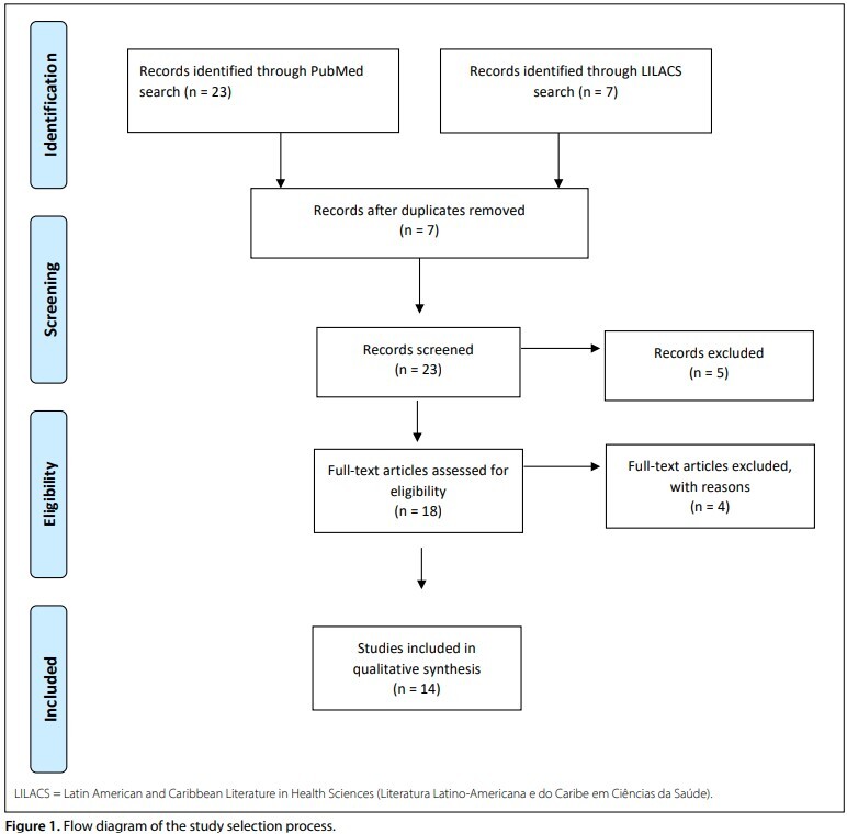 Figure 1
| Database | Search strategy | Papers found |
|---|---|---|
| MEDLINE (via PubMed) | (“artificial intelligence”) and (“ophthalmology”) and (“cardiovascular disease”) | 17 |
| MEDLINE (via PubMed) | (“artificial intelligence”) and (“ophthalmology”) and (“stroke”) | 0 |
| LILACS (via Biblioteca Virtual em Saúde) | (“artificial intelligence”) and (“ophthalmology”) and (“cardiovascular disease”) | 7 |
| LILACS (via Biblioteca Virtual em Saúde) | (“artificial intelligence”) and (“ophthalmology”) and (“stroke”) | 0 |
| MEDLINE (via PubMed) | (“prediction”) and (“biomarker”) and (“stroke”) and (“retina”) | 2 |
| LILACS (via Biblioteca Virtual em Saúde) | (“prediction”) and (“biomarker”) and (“stroke”) and (“retina”) | 0 |
| MEDLINE (via PubMed) | (“prediction”) and (“biomarker”) and (“cardiovascular disease”) and (“retina”) | 4 |
| LILACS (via Biblioteca Virtual em Saúde) | (“prediction”) and (“biomarker”) and (“cardiovascular disease”) and (“retina”) | 0 |
| Article/Year | Pathology/Evaluated factor | Main Findings / Metrics |
|---|---|---|
| Poplin et al.
| Cardiovascular disease/ 284,335 patients | Gender (AUC = 0.97), smoking status (AUC = 0.71), major adverse cardiac events (AUC = 0.70) |
| Cheung CY et al.
| Cardiovascular disease/ 70,000 retinal photographs | Blood pressure, BMI, and cholesterol level correlated with vascular changes (r = 0.82–0.95) |
| Rim et al.
| Cardiovascular disease/ 216 152 retinal photographs | Coronary artery calcium (AUC = 0.742; 95%CI = 0.732–0.753) |
| Arnould et al.
| Cardiovascular disease/ 237 patiens-OCTa | SCP density associated with cardiovascular risk profile (OR = 1.06) |
| Chang et al.
| Cardiovascular disease/ 15,408 retinal photographs | Prediction of atherosclerosis (AUC = 0.713) |
| Song et al.
| Cardiovascular disease/ 44,184 retinal photographs | AUC = 82.3% (95%CI = 79.5%–85.0%) |
| Zhang et al.
| Cardiovascular disease/ 1,222 fundus images | AUC = 0.766 (hypertension), AUC = 0.703 (dyslipidemia) |
| Zekavat et al.
| Cardiovascular disease/ 97,895 retinal fundus images | Retinal vasculature as biomarker for cardiometabolic and ocular disease |
| Tseng et al.
| Cardiovascular disease/ 48,260 patients | Reti-CVD associated with increased CVD risk (HR = 1.41; 95% CI) |
| Zhu et al.
| Stroke/80,169 fundus images | Stroke risk increased by 4% per unit retinal age gap (HR = 1.04; 95%CI = 1.00–1.08) |
| Nosinovic et al.
| Cardiovascular disease/ 129,236 retinal photographs | Sensitivity = 0.70, specificity = 0.55 for predicting cardiovascular mortality |
| Dai et al.
| Cardiovascular disease/ 2,012 retinal photographs | AUC = 0.6506 for hypertension prediction via heat map algorithms |
| Yi et al.
| Cardiovascular disease/ 48,260 participants | Sensitivity = 82.7%, specificity = 84.0% for CVD risk prediction |
| Rudnicka et al.
| Cardiovascular disease and stroke/ 65,144 participants | Algorithm outperformed or equaled FRS for MI and stroke |
Peer Reviews
No public reviews on file for this paper yet. If you reviewed it on a platform where reviews are public (OpenReview, ICLR, NeurIPS, ICML), you can paste yours below so the community can read it here.
Videos
No videos yet. Explain this paper in a talk, walkthrough, or lecture? Add one.
Taxonomy
TopicsRetinal Imaging and Analysis · Retinal and Optic Conditions · Acute Ischemic Stroke Management
INTRODUCTION
Cardiovascular diseases (CVDs) are among the leading causes of death worldwide, resulting in significant socioeconomic losses.^ 1 ^ Controlling risk factors for CVDs can reduce mortality rates and healthcare-related expenditures.
The eyes serve as a window into cardiovascular health, functioning as a valuable non-invasive tool for studying and monitoring CVDs. Studies have shown that alterations in microcirculation can predict macroscopic changes associated with CVD. Retinal arteries and veins measure approximately 150–200 micrometers, and vascular narrowing is an indicator of chronic hypertension.^ 2 ^ The narrowing of retinal arteries has already been documented as a predictor of CVD and stroke.^ 3,4,5 ^
OBJECTIVE
This article presents a review of the application of artificial intelligence in identifying biomarkers for CVD and stroke.
METHODS
A literature search was conducted on the main applications of artificial intelligence in ophthalmology, using the keywords “artificial intelligence,” “prediction,” “biomarker,” “cardiovascular disease,” “retina,” and “stroke,” covering the period from January 1, 2018, to July 3, 2023. The Medical Literature Analysis and Retrieval System Online (MEDLINE) database (via PubMed) and Latin American and Caribbean Literature in Health Sciences (LILACS) (via the Virtual Health Library) were used to identify relevant articles. Articles discussing the potential automated clinical applications of artificial intelligence technologies were selected and reviewed by the authors. A summary of the selected articles is provided below, and the details of the search strategy are shown in Table 1.
An initial screening of articles was performed based on the abstracts. At this stage, studies that did not meet the inclusion criteria were excluded.
Subsequently, a second-level screening excluded articles that were not sufficiently comparable. Finally, the selected studies were grouped by similar characteristics to derive conclusions and new insights based on the gathered data.
RESULTS
After screening titles and abstracts, removing duplicates, and reviewing citations, 14 studies were deemed eligible for critical analysis. The article selection process is illustrated in Figure 1.
Flow diagram of the study selection process.
Cardiovascular disease
Algorithms evaluating fundus image
Oculomics is a promising tool that adds value to the diagnosis and monitoring of CVDs, which are among the leading causes of mortality worldwide.^ 6 ^ For the analysis of ophthalmologic alterations in patients with CVD, the primary algorithms used include Integrative Vessel Analysis, Singapore I Vessel Assessment, and Vascular Assessment and Measurement Platform for Retinal Imaging. These algorithms enable the evaluation of microvascular parameters of arterioles, venules, and the optic nerve head. They also assess vascular tortuosity and branching angles.^ 6,7,8 ^
Poplin et al. developed an algorithm to assess cardiovascular risk. They analyzed 284,335 patients and were able to predict cardiovascular risk factors using retinal imaging—such as gender (area under the curve [AUC] = 0.97), smoking status (AUC = 0.71), and major adverse cardiac events (AUC = 0.70). The study demonstrated that the trained deep learning models utilized anatomical features, such as the optic disc and blood vessels, to generate each prediction (Table 2).^ 9 ^
Cheung CY et al.^ 10 ^ analyzed 70,000 fundus images and correlated cardiovascular risk factors with retinal vascular alterations, including blood pressure, body mass index, and cholesterol levels, reporting correlation coefficients ranging from 0.82 to 0.95. The models performed comparably to experts in assessing the caliber of retinal vessels and predicting cardiovascular risk factors, including blood pressure, cholesterol levels, and glycated hemoglobin.
Rim et al. used 216,152 fundus images to predict and stratify CVD risk by identifying the presence of coronary artery calcium, reporting an AUC of 0.742 (95% confidence interval [CI]: 0.732–0.753). These findings suggest that algorithms analyzing fundus images can serve as predictors of coronary artery calcium levels, particularly in resource-limited settings.^ 11 ^
Zhu et al. analyzed 80,169 fundus images from 46,969 participants and found no association between retinal image–based age estimation and CVD risk. However, the study demonstrated that retinal age difference may be a potential biomarker correlated with mortality risk.^ 12 ^
Chang et al.^ 13 ^ analyzed 15,408 fundus images to predict carotid artery atherosclerosis and achieved an AUC of 0.713, demonstrating the potential to detect atherosclerosis using retinal images.
Song et al. were able to assess coronary artery calcium accumulation risk by analyzing 44,184 fundus images. The study reported AUCs of 82.3% (79.5%–85.0%) and 83.2% (80.2%–86.3%) using unilateral and bilateral fundus images, respectively. Performance improved when bilateral images were used. The study primarily analyzed the retinal vessels and the fovea.^ 14 ^
Zang et al. analyzed 1,222 fundus images and developed an algorithm with an AUC of 0.766 for predicting hypertension and 0.703 for predicting dyslipidemia—both key risk factors for CVD. The algorithms demonstrated the feasibility of predicting dyslipidemia, hypertension, diabetes, and other cardiovascular conditions.^ 15 ^
Zekavat et al.^ 16 ^ analyzed 97,895 retinal fundus images and assessed retinal vessels by calculating vascular density and fractal dimension as measures of vascular branching complexity. They proposed that retinal vasculature may serve as a biomarker for future cardiometabolic and ocular diseases.
Tseng et al.^ 17 ^ examined 48,260 patients without a history of CVD using retinography over an 11-year follow-up period, during which 6.3% of patients developed CVD. The identified biomarker, Reti-CVD, was associated with an increased risk of CVD (adjusted hazard ratio [HR]: 1.41; 95%CI: not specified). Reti-CVD may be useful in identifying individuals with a ≥ 10% 10-year CVD risk who could benefit from early preventive interventions. It could assist in accurately stratifying individuals at higher cardiovascular risk.
Nosinovic et al.^ 18 ^ analyzed 129,236 retinal photographs from 40,480 participants and demonstrated the ability to predict CVD–related mortality, achieving a sensitivity of 0.70 and specificity of 0.55.
Dai et al.^ 19 ^ analyzed 2,012 retinal photographs and reported an AUC of 0.6506 in developing heatmaps through algorithmic analysis to detect hypertension, a significant CVD risk factor. The study identified an association between systemic arterial hypertension and vessel branching.
Yi et al.^ 20 ^ evaluated 48,260 participants, and their algorithm analyzed retinography images to classify individuals at risk of developing CVD, achieving a sensitivity of 82.7% and specificity of 84.0%. The retinal photographic biomarker (Reti-CVD) successfully identified five individuals at intermediate and high risk for CVD.
Algorithms evaluating optical coherence tomography
The use of optical coherence tomography angiography (OCT-A) to measure superficial retinal capillary plexus (SCP) vascular density in 237 patients identified that patients with lower vascular density had a higher risk of hypertension, diabetes, and CVD (odds ratio [OR]: 1.06; 95%CI not fully reported).^ 21 ^ OCT-A is expected to play a critical role in the discovery of new biomarkers for CVD in the future.
Stroke
Zhu et al.^ 22 ^ analyzed 80,169 fundus images from 46,969 participants. The algorithm processed the images and calculated the retinal age gap, which was associated with a 4% increase in stroke risk (HR = 1.04; 95%CI = 1.00–1.08; P = 0.029). The study concluded that the retinal age gap was significantly associated with incident stroke, suggesting its potential as a predictive biomarker for stroke risk.
Rudnicka et al.^ 23 ^ analyzed 65,144 participants and developed an algorithm that outperformed the Framingham risk scores in predicting myocardial infarction and stroke. The algorithm offers a non-invasive biomarker for vascular risk assessment, eliminating the need for blood tests or blood pressure measurements.
DISCUSSION
The development of algorithms for the diagnosis of ophthalmologic diseases requires careful consideration to ensure both the effectiveness and safety of the system. It is essential that algorithms be designed and trained on a diverse set of images representative of the population to which they will be applied. Different ethnic groups, age ranges, genders, and ocular conditions must be included to ensure that the algorithm can generalize its diagnoses accurately and equitably. Moreover, the algorithm must be based on robust and current clinical evidence. The data used for training should originate from reputable sources, such as peer-reviewed scientific studies and clinical datasets from established medical institutions. Algorithms should also be tested on independent datasets (validation sets) to confirm their effectiveness in real-world scenarios.
Furthermore, it is important to ensure that the algorithm can be generalized across different medical centers and ophthalmic equipment. Although an algorithm may initially be developed to diagnose a specific disease, expanding its application to a broader range of ocular conditions may enhance its utility for both healthcare professionals and patients. Transparency is essential for promoting trust in algorithmic outputs. Algorithms should be designed to provide interpretable information about how diagnostic conclusions are reached, enabling clinicians to understand and verify the results. Finally, algorithms must be continuously updated as new clinical evidence emerges. There should be a defined mechanism for implementing updates to improve diagnostic accuracy and maintain clinical relevance.
The analysis of vascular tortuosity and segmentation of retinal vessels can serve as a non-invasive biomarker in the study of CVD. Existing factors reported in the literature have demonstrated the ability to predict stroke risk with a maximum accuracy of approximately 60%.^ 5 ^ Consequently, review studies play an important role in generating new evidence that supports the development of research lines in underexplored areas.
Ethical and legal considerations are fundamental during data analysis and algorithm development, particularly in medical applications such as the diagnosis of ocular diseases. It is essential to ensure that patient information and images used for algorithm training are collected and stored securely, in compliance with applicable data protection laws and regulations. Data should be anonymized whenever possible to prevent the identification of individual patients. If training data include personal patient information, obtaining informed consent is critical prior to its collection and use for research and development purposes. Furthermore, awareness of potential bias in training data is important, as such bias may lead to inequitable or inaccurate algorithmic outcomes.
Training datasets must be carefully curated, and physicians should be able to understand how the artificial intelligence system generated a diagnosis in order to have confidence in the results and enable independent validation by other researchers.
The use of federated learning reduces ethical concerns related to the retention of training data by enabling algorithms to be trained locally by users, thereby improving performance and mitigating training sample bias. This approach leverages user-side processing, facilitating decentralized algorithm development while enhancing data privacy. Sharing algorithms across research groups should be encouraged to accelerate the development of novel biomarkers.^ 24 ^
The application of artificial intelligence in ophthalmology can contribute not only to a better understanding of the pathophysiology of CVDs through the identification of novel biomarkers but also to the diagnosis and monitoring of these diseases in settings with limited healthcare resources. As non-invasive techniques, such approaches gain value when considered alongside established cardiovascular risk factors.
Comparing the performance of different algorithms for detecting CVD or stroke is challenging due to variations in population samples and inconsistency in evaluation parameters. This underscores the need for studies that include external validation of algorithms.
Future directions, strengths, and limitations
Algorithms that analyze fundus images may be affected by variations in brightness, focus, and contrast during training. Additional prospective and randomized studies are necessary to establish novel biomarkers for monitoring CVD.
Narrative reviews are valuable for identifying knowledge gaps and guiding new lines of research aimed at developing biomarkers for the diagnosis of CVD. Future studies should incorporate multimodal analysis, combining the use of OCT, OCT-A, and fundus image analysis to enhance understanding of CVD pathophysiology. OCT-A enables the evaluation of different vascular plexuses of the retina, including vessel density and perfusion. It is also important that future research distinguish between biomarkers associated with ischemic and hemorrhagic stroke.
External validation of developed algorithms should be consistently encouraged to assess performance across geographically and demographically distinct populations.
Most existing algorithms have been applied to two-dimensional retinal images. Training with multimodal datasets may facilitate the development of three-dimensional image analysis, thereby supporting the development of more accurate biomarkers.
CONCLUSION
The use of artificial intelligence has advanced understanding of the pathophysiology of CVD and has contributed to the identification of novel non-invasive biomarkers through the analysis of ophthalmologic images.
The reference list from the paper itself. Each links out to its DOI / PubMed record.
- 1Dagenais GR Leong DP Rangarajan S Variations in common diseases, hospital admissions, and deaths in middle-aged adults in 21 countries from five continents (PURE): a prospective cohort study Lancet 20203951022678579410.1016/s 0140-673631492501 · doi ↗ · pubmed ↗
- 2Wang L Wong TY Sharrett AR Relationship between retinal arteriolar narrowing and myocardial perfusion: multi-ethnic study of atherosclerosis Hypertension 20085111192610.1161/hypertensionaha.107.09834317998474 · doi ↗ · pubmed ↗
- 3Wong TY Klein R Sharrett AR Retinal arteriolar narrowing and risk of coronary heart disease in men and women The Atherosclerosis Risk in Communities Study. JAMA 200228791153910.1001/jama.287.9.115311879113 · doi ↗ · pubmed ↗
- 4Wang JJ Liew G Klein R Retinal vessel diameter and cardiovascular mortality: pooled data analysis from two older populations Eur Heart J 2007281619849210.1093/eurheartj/ehm 22117626032 · doi ↗ · pubmed ↗
- 5Cheung CY Tay WT Ikram MK Retinal microvascular changes and risk of stroke: the Singapore Malay Eye Study Stroke 20134492402810.1093/eurheartj/ehm 22123868266 · doi ↗ · pubmed ↗
- 6Parr JC Spears GF Mathematic relationships between the width of a retinal artery and the widths of its branches Am J Ophthalmol 19747744788310.1016/0002-93944819452 · doi ↗ · pubmed ↗
- 7Parr JC Spears GF General caliber of the retinal arteries expressed as the equivalent width of the central retinal artery Am J Ophthalmol 1974774472710.1016/0002-93944819451 · doi ↗ · pubmed ↗
- 8Hubbard LD Brothers RJ King WN Methods for evaluation of retinal microvascular abnormalities associated with hypertension/sclerosis in the Atherosclerosis Risk in Communities Study Ophthalmology 19991061222698010.1016/s 0161-642010599656 · doi ↗ · pubmed ↗