Detective flow imaging, directional enhanced blood flow imaging, and contrast-enhanced harmonic endoscopic ultrasound in pancreatic solid lesions
Filippo Antonini, Durante Donnarumma, Tiziana Buono

Abstract
Genes, proteins, chemicals, diseases, species, mutations and cell lines named across the full text — each resolved to its canonical identifier and authoritative record.
Click any figure to enlarge with its caption.
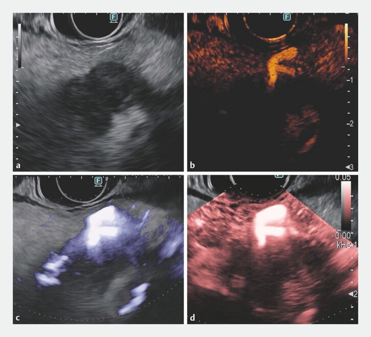 Fig. 1
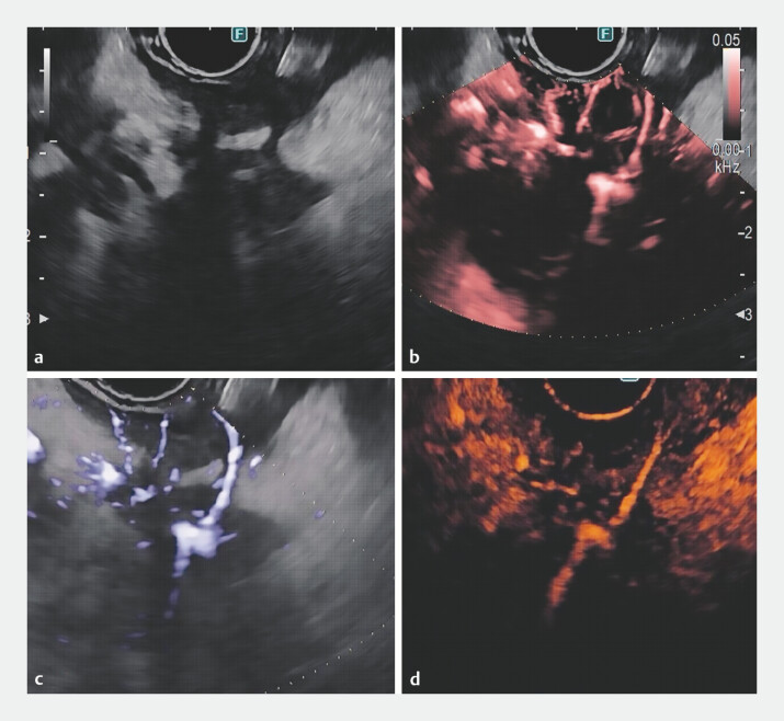 Fig. 2
Peer Reviews
No public reviews on file for this paper yet. If you reviewed it on a platform where reviews are public (OpenReview, ICLR, NeurIPS, ICML), you can paste yours below so the community can read it here.
Videos
No videos yet. Explain this paper in a talk, walkthrough, or lecture? Add one.
Taxonomy
TopicsPancreatic and Hepatic Oncology Research · Neuroendocrine Tumor Research Advances · MRI in cancer diagnosis
Contrast-enhanced harmonic endoscopic ultrasound (CH-EUS) is known for its superior diagnostic accuracy for solid pancreatic lesions (SPLs), primarily due to its enhanced visualization of perfusion patterns 1 . However, it may not be suitable for all patients and relies on the use of expensive contrast agents. In contrast, directional enhanced blood flow imaging (eFLOW) and the more recent detective flow imaging endoscopic ultrasound (DFI-EUS) are innovative imaging techniques that improve the visualization of fine vascular structures in various tissues, all without the need for contrast agents 2 3 .
We present a brief series of cases of both malignant and benign SPLs evaluated through eFLOW and DFI-EUS, with a comparative analysis using CH-EUS ( Fig. 1 , Fig. 2 , Video 1 ). Notably, this series includes a rare case highlighting the application of DFI in a patient diagnosed with autoimmune pancreatitis.
A case of pancreatic cancer: a B-mode imaging reveals a hypoechoic lesion. On ( b ) CH, ( c ) directional eFLOW, and ( d ) DFI endoscopic ultrasound, the tumor consistently shows a hypovascular pattern with central areas of increased blood flow. Abbreviations: CH, contrast-enhanced harmonic; DFI, detective flow imaging; eFLOW, enhanced blood flow imaging.
A case of degenerated IPMN: a B-mode imaging shows a hypoechoic area with irregular margins. On ( b ) DFI, ( c ) directional eFLOW, and ( d ) CH endoscopic ultrasound, the characteristic hypo-enhancement of pancreatic cancer is observed, along with a linear central vessel. A diagnostic EUS-guided fine needle biopsy was performed on the hypoechoic area, carefully avoiding the vessel. Abbreviations: CH, contrast-enhanced harmonic; DFI, detective flow imaging; eFLOW, enhanced blood flow imaging; EUS, endoscopic ultrasound; IPMN, intraductal papillary mucinous neoplasm.
Solid pancreatic lesions evaluated through directional enhanced blood flow imaging (eFLOW) and detective flow imaging (DFI), with a comparative analysis using contrast-enhanced harmonic endoscopic ultrasound (CH-EUS).Video 1
CH-EUS, DFI-EUS, and eFLOW can provide complementary insights into the vascular characteristics of the lesions. All three techniques tend to reveal similar findings in terms of vascular patterns: in pancreatic adenocarcinoma, these imaging modalities typically show a hypovascular pattern, reflecting the poor blood supply and disorganized blood vessels that are characteristic of malignancy, often associated with areas of increased blood flow or neoangiogenesis within the tumor.
While CH-EUS typically offers the most comprehensive analysis of blood perfusion in SPLs, established criteria for classifying vessels with DFI-EUS and eFLOW are still lacking. However, using these modalities in a complementary manner can significantly enhance the differential diagnosis of pancreatic lesions. Both eFLOW and DFI-EUS provide valuable alternatives, especially for patients who are unable to receive contrast agents.
Endoscopy_UCTN_Code_TTT_1AS_2AD
The reference list from the paper itself. Each links out to its DOI / PubMed record.
- 1Otsuka Y Kamata KA review of contrast-enhanced harmonic endoscopic ultrasonography for pancreatic solid tumors [Epub ahead of print. Erratum in: J Med Ultrason (2001). 2024. doi:10.1007/s 10396–024–01435-x. PMID: 37584780]J Med Ultrason (2001)202310.1007/s 10396-023-01346-337584780 · doi ↗ · pubmed ↗
- 2Miwa H Sugimori K Yonei S Differential Diagnosis of Solid Pancreatic Lesions Using Detective Flow Imaging Endoscopic Ultrasonography Diagnostics (Basel)20241488210.3390/diagnostics 1409088238732296 PMC 11082975 · doi ↗ · pubmed ↗
- 3Mulqui MV Caillol F Ratone JP Detective flow imaging versus contrast-enhanced EUS in solid pancreatic lesions Endosc Ultrasound 20241324825210.1097/eus.000000000000007639318752 PMC 11419480 · doi ↗ · pubmed ↗