A novel biopsy forceps assisted the removal of a deeply displaced pancreatic duct stent with pancreatic duct stenosis under the guidance of a peroral choledochoscope
Jiao Tian, Yankun Hou, Tingting Yu, Lichao Zhang, Guoying Wang, Senlin Hou

Abstract
Genes, proteins, chemicals, diseases, species, mutations and cell lines named across the full text — each resolved to its canonical identifier and authoritative record.
Click any figure to enlarge with its caption.
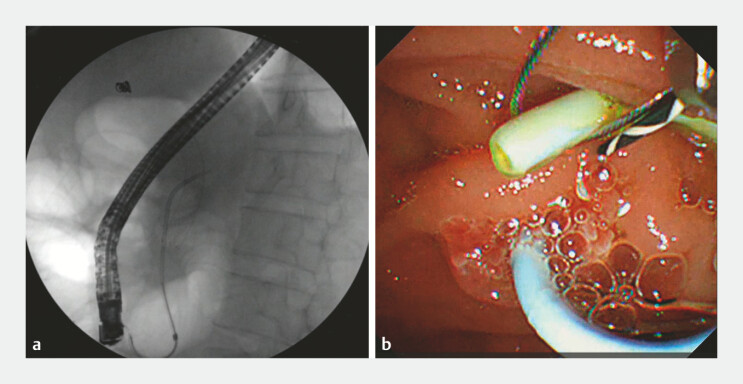 Fig. 1
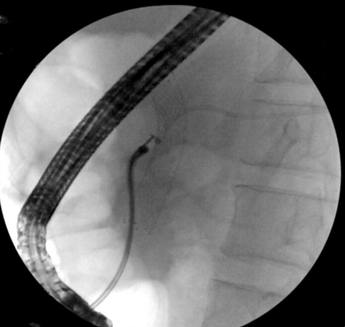 Fig. 2
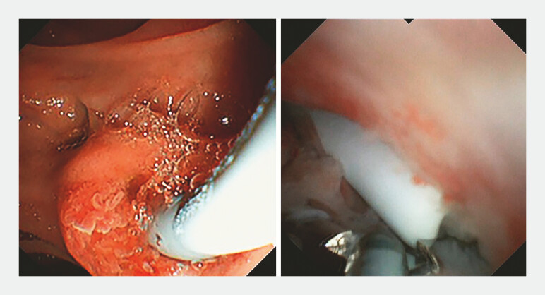 Fig. 3
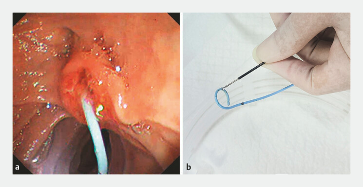 Fig. 4
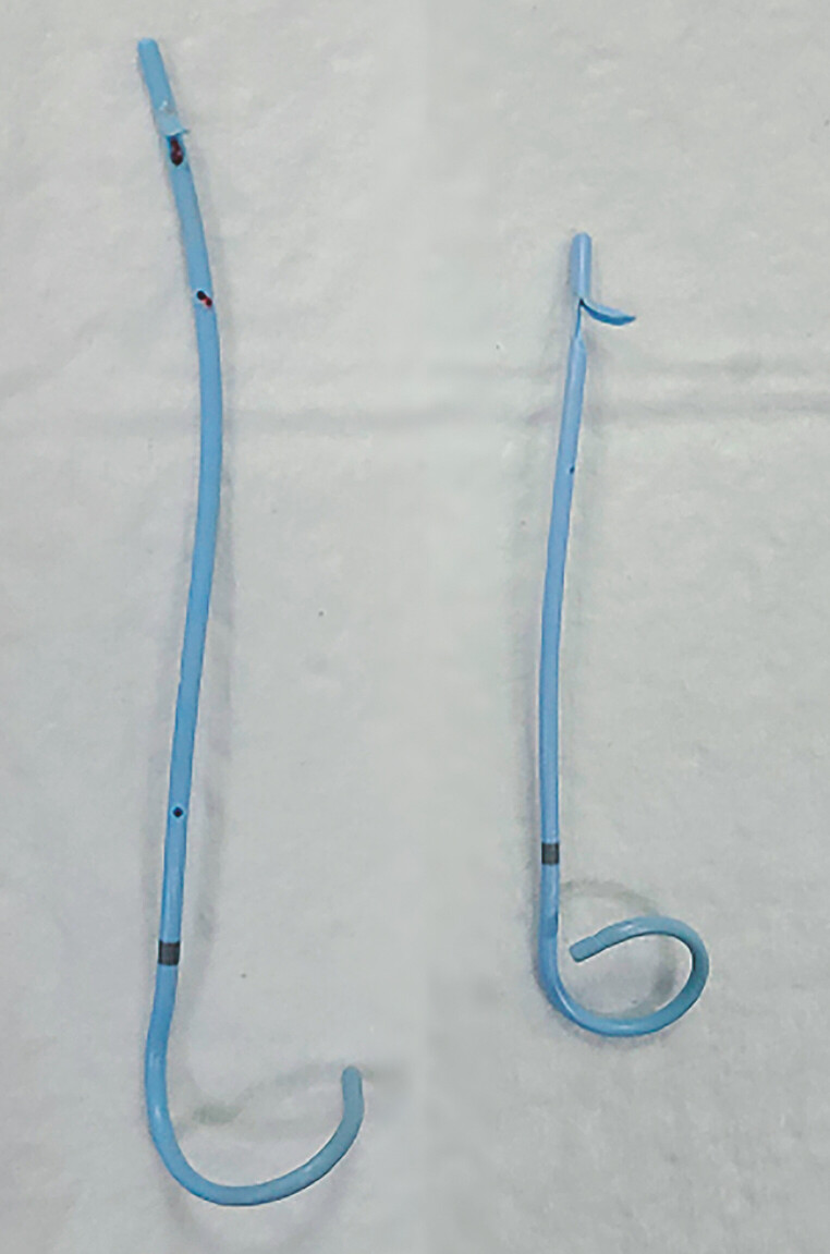 Fig. 5
Peer Reviews
No public reviews on file for this paper yet. If you reviewed it on a platform where reviews are public (OpenReview, ICLR, NeurIPS, ICML), you can paste yours below so the community can read it here.
Videos
No videos yet. Explain this paper in a talk, walkthrough, or lecture? Add one.
Taxonomy
TopicsEsophageal and GI Pathology · Pediatric Hepatobiliary Diseases and Treatments · Gallbladder and Bile Duct Disorders
Endoscopic removal of displaced pancreatic duct stents was challenging, especially in patients with pancreatic duct stenosis 1 . We present a novel biopsy forceps for the assisted removal of a deeply displaced pancreatic duct stent with pancreatic duct stenosis.
A 42-year-old man underwent a routine endoscopic retrograde cholangiography (ERCP) for chronic pancreatitis. Unfortunately, the pancreatic duct stent was displaced to the tail of the pancreas, and because the stent with the stenosis of the pancreatic duct could not be removed, a new pancreatic duct stent was inserted. The patient was referred to our center, where we removed the first stent with a snare ( Fig. 1 a, b ). Then into the peroral choledochoscope ( Fig. 2 ), a novel biopsy forceps was applied under direct vision after the pancreatic duct stent was located (SpyBite Max, Boston Scientific Corporation) grab the displaced stent and pull it to the duodenal papilla ( Fig. 3 ). It is then removed with a snare and replaced with a pancreatic duct stent ( Fig. 4 a, b , Fig. 5 , Video 1 ). The patient had no significant postoperative discomfort and was discharged 3 days after surgery.
a The displaced pancreatic duct stent is visible below the X-ray line. b Remove the first bracket with a snare.
Enter the peroral choledochoscope to assist in removing the pancreatic duct stent displaced into the tail of the pancreas.
The displaced pancreatic duct stent was pulled to the duodenal papilla by biopsy forceps under the direct vision of peroral choledochoscope.
The pancreatic duct stent was pulled to the duodenal papilla and removed with a snare.
The pancreatic duct stent was removed from the body.
Novel biopsy forceps assisted the removal of a deeply displaced pancreatic duct stent with pancreatic duct stenosis under the guidance of peroral choledochoscope.Video 1
The removal of pancreatic duct stents with deep displacement has different methods in different centers 2 3 . We used a newly marketed biopsy forceps that has more rodent-shaped jaws than conventional biopsy forceps for a stronger grasp. At the same time, the biopsy forceps can be fine-adjusted repeatedly, making it easier to operate in narrow spaces. The application of the biopsy forceps provides a new way for the treatment of such patients, making it possible to remove foreign bodies in the pancreatic duct under direct vision.
Endoscopy_UCTN_Code_CPL_1AK_2AD
The reference list from the paper itself. Each links out to its DOI / PubMed record.
- 1Cai YL Wang F Li ZS Endotherapy for the Proximal Migration of Pancreatic Stents: A Systematic Review Pancreas 202453 e 694e 69910.1097/MPA.000000000000235438696448 · doi ↗ · pubmed ↗
- 2Ng WK Tan QR Punamiya SJ Novel method to remove deeply migrated pancreatic duct stent Endoscopy 202254 E 748E 74910.1055/a-1792-246935359016 · doi ↗ · pubmed ↗
- 3Kato A Natsume M Yoshida M Endoscopic tapered sheath-assisted removal of a proximally migrated pancreatic stent Endoscopy 202254 E 767E 76810.1055/a-1792-295535359015 PMC 9735239 · doi ↗ · pubmed ↗