Underwater resection of cecal submucosal tumors using cap, clip, and snare assistance
Chang-en Liu, Yan Li

Abstract
Genes, proteins, chemicals, diseases, species, mutations and cell lines named across the full text — each resolved to its canonical identifier and authoritative record.
Click any figure to enlarge with its caption.
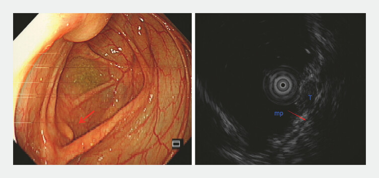 Fig. 1
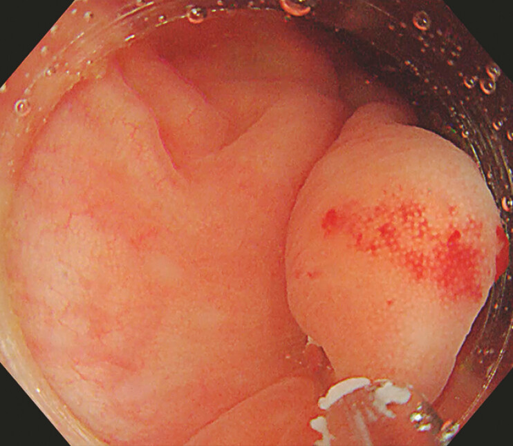 Fig. 2
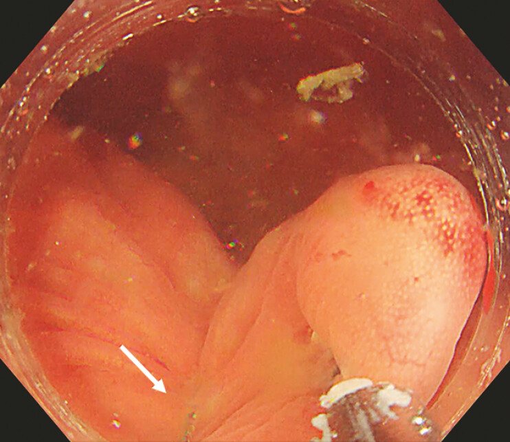 Fig. 3
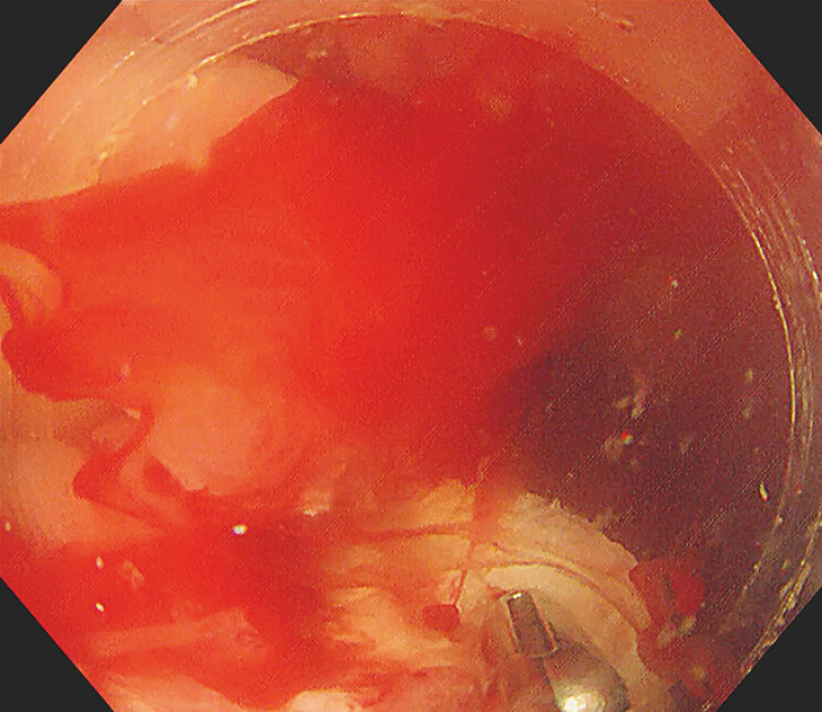 Fig. 4
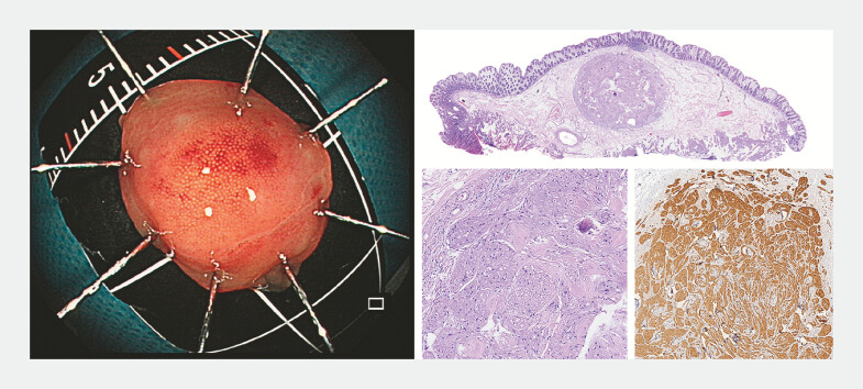 Fig. 5
- —Tianjin Key Medical Discipline (Specialty) Construction Project
Peer Reviews
No public reviews on file for this paper yet. If you reviewed it on a platform where reviews are public (OpenReview, ICLR, NeurIPS, ICML), you can paste yours below so the community can read it here.
Videos
No videos yet. Explain this paper in a talk, walkthrough, or lecture? Add one.
Taxonomy
TopicsGastrointestinal Tumor Research and Treatment · Salivary Gland Tumors Diagnosis and Treatment · Tracheal and airway disorders
Clip-and-snare assisted endoscopic mucosal resection (CS-EMR) is a safe and effective treatment for small rectal neuroendocrine tumors (NETs) 1 . Following this approach, we applied the underwater method, U-CCS-EMR, for resecting submucosal lesions in the cecum.
A 42-year-old asymptomatic woman was identified with a 5 mm yellow, elevated submucosal lesion situated at the cecum during a routine colonoscopy. Ultrasonography (12 MHz) confirmed the lesionʼs location within the submucosal layer ( Fig. 1 ). Endoscopic resection was requested.
Yellow submucosal lesion (red arrow) confirmed by ultrasound to be located in the submucosal layer.
A transparent cap-covered single-channel colonoscopy, along with a pre-anchored snare, was inserted into the ileocecal region to target the lesion. The lesion, in a low-lying position, was obscured by residual fecal water, which significantly hindered both observation and treatment. Consequently, we opted for an innovative underwater treatment method ( Video 1 ). A clip was introduced via the working channel of the endoscope to secure the mucosa adjacent to the lesion ( Fig. 2 ). When the lesion and surrounding tissues were well lifted by the clip, the snare was released from the transparent cap and completely enveloped the root of the lesion ( Fig. 3 ). The lesion was entirely resected en bloc, leaving a clean surgical wound. The wound was promptly closed using clips. The patient was placed on a 24-hour fast following the procedure and was discharged in good condition 2 days later with no complications.
Endoscopic removal of the cecal submucosal tumor.Video 1
A clip was placed via the endoscope to secure the mucosa under the lesion.
Release the snare and position it at the base of the pseudo-pedicle.
U-CCS-EMR is a promising treatment for cecal submucosal tumors. It provides a clear view throughout the procedure, especially during bleeding ( Fig. 4 ). It also preserves the submucosal layer’s natural laxity, aiding in the formation of a pseudo-long pedicle and ensuring a negative resection margin. Postoperative pathology revealed the lesion to be a granular cell tumor ( Fig. 5 ). Additionally, it reduces thermal damage to the muscle layer, lowering the risk of delayed perforation. Our experience shows that underwater resection with cap, clip, and snare assistance is both safe and effective.
Underwater view: clearly visible low-position lesion and mini arterial bleeding point.
The lesion was resected en bloc and histopathology indicated granular cell tumor: HE, HE (20×), and S100 (+).
Endoscopy_UCTN_Code_TTT_1AQ_2AD_3AF
The reference list from the paper itself. Each links out to its DOI / PubMed record.