Right extrapleural hematoma due to thoracic trauma. The extrapleural fat sign
Raquel García-Latorre, Luis Gorospe, Abel González-Huete

Abstract
Genes, proteins, chemicals, diseases, species, mutations and cell lines named across the full text — each resolved to its canonical identifier and authoritative record.
Click any figure to enlarge with its caption.
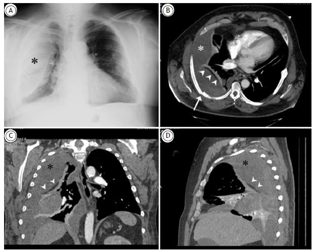 Figure 1
Peer Reviews
No public reviews on file for this paper yet. If you reviewed it on a platform where reviews are public (OpenReview, ICLR, NeurIPS, ICML), you can paste yours below so the community can read it here.
Videos
No videos yet. Explain this paper in a talk, walkthrough, or lecture? Add one.
Taxonomy
TopicsTrauma Management and Diagnosis · Case Reports on Hematomas · Congenital Diaphragmatic Hernia Studies
We report the case of a 54-year-old male presenting to the emergency department with dyspnea and right-sided chest pain following recent thoracic trauma. He had no significant medical history but showed progressive anemia.
A chest X-ray (Figure 1A) revealed right-sided rib fractures and an extensive ipsilateral opacity with extrapulmonary morphology. Chest CT scan (Figures 1B-D) showed a loculated, biconvex collection on the right side, with dependent areas of bleeding, separated from the lung parenchyma by a thin fat-density line (the extrapleural fat sign). These findings confirmed an extrapleural hematoma. The hematoma was successfully drained via a chest tube, leading to clinical improvement.
Figure 1(A) Posteroanterior chest X-ray showing right rib fractures and a large extrapulmonary opacity (asterisk). Axial (B), coronal (C), and sagittal (D) reconstructions of chest CT scan (mediastinal window) reveal rib fractures (arrow) and a large extrapulmonary collection (asterisks) separated from the atelectatic lung parenchyma by a linear fat-density image (arrowheads) corresponding to the extrapleural fat sign, consistent with an extrapleural hematoma.
Extrapleural hematomas are rare, occurring in 7.1% of thoracic trauma cases. They result from bleeding between the parietal pleura and endothoracic fascia and are often associated with rib fractures, hemothorax, pneumothorax, and pulmonary contusions.1
The extrapleural fat sign, seen on CT, is a linear fat-density line separating the pulmonary parenchyma from extrapleural lesions. It corresponds to extrapleural fat thickened and medially displaced in extrapleural pathologies.2
Recognizing this sign is critical to differentiating extrapleural hematomas from hemothorax, as their management and complications differ.1 ^,^ 2
The reference list from the paper itself. Each links out to its DOI / PubMed record.
- 1Shankar T Ameena Ms S Nagasubramanyam V Meena R Sasidharan P Extrapleural Hematoma A Rare Sequalae of Thoracic Trauma Cureus 2024169 e 7050610.7759/cureus.7050639479078 PMC 11524070 · doi ↗ · pubmed ↗
- 2Chung JH Carr RB Stern EJ Extrapleural hematomas imaging appearance, classification, and clinical significance J Thorac Imaging 201126321822310.1097/RTI.0b 013e 3181 ebeaba 20818277 · doi ↗ · pubmed ↗