Epithelioid Sarcoma Mimicking Mycotic Granulomatosis
Yoichi Kaneuchi, Michiyuki Hakozaki, Takuya Nikaido, Yoshihiro Matsumoto

Abstract
Genes, proteins, chemicals, diseases, species, mutations and cell lines named across the full text — each resolved to its canonical identifier and authoritative record.
Click any figure to enlarge with its caption.
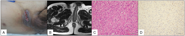 Figure 1
Peer Reviews
No public reviews on file for this paper yet. If you reviewed it on a platform where reviews are public (OpenReview, ICLR, NeurIPS, ICML), you can paste yours below so the community can read it here.
Videos
No videos yet. Explain this paper in a talk, walkthrough, or lecture? Add one.
Taxonomy
TopicsInfectious Diseases and Mycology · Cutaneous lymphoproliferative disorders research · Fungal Infections and Studies
A 51-year-old man was referred to our hospital with a 2-year history of a slow-growing refractory skin ulcer on his right groin. The physical examination showed a painless ulcer with firm edges, measuring 6 cm × 2 cm (Figure 1A), but no inguinal lymphadenopathy. The laboratory examination revealed a white cell count of 5,500/mL (reference range, 3,300-8,600/mL) and the C-reactive protein level of 0.12 mg/L (reference range, <0.30 mg/L). Magnetic resonance imaging (MRI) showed an infiltrative superficial soft-tissue mass appearing homogenous and low intensity on T1-weighted images (Figure 1B), heterogeneous with low and iso-intensity on T2-weighted images, and heterogenous on gadolinium-enhanced T1-weighted fat-suppression images. Mycotic granulomatosis was clinically suggested, and a skin biopsy was performed. Bacterial, fungal, and mycobacterium cultures were all negative. The histopathological examination after a second biopsy revealed large, ovoid epithelioid cells with rich eosinophilic cytoplasm (Figure 1C). The immunohistochemical examination revealed that the tumor cells were positive for pan-cytokeratin, CD34, and Friend leukemia integration 1 transcription factor but negative for BAF47/integrase interactor 1 (Figure 1D), leading to the diagnosis of epithelioid sarcoma, which is an extremely rare malignant soft-tissue tumor ^(1)^. The patient underwent a wide resection with a right orchiectomy. Antibiotics were administered long term, but owing to a surgical site infection at one month postoperatively, debridement and placement of antibiotic-loaded cement beads were performed, causing the resolution of the infection. Truncal computed tomography and pelvic MRI were performed every 3 months until 2 years postoperatively, and imaging examinations were continued every 4 months thereafter. At 4 years postoperatively, there is no evidence of metastasis or recurrence. Localized epithelioid sarcoma shows a relatively high overall survival rate of 62%-88%, whereas the rate decreases 24% in patients with metastasis ^(2)^. Lymph node dissemination and metastasis rates for localized epithelioid sarcoma are 34%-52% and 33%, respectively. Epithelioid sarcoma is extremely rare but should be considered as a differential diagnosis when a skin ulcer does not heal over a long period.
Article Information
Conflicts of Interest
None
The reference list from the paper itself. Each links out to its DOI / PubMed record.