Characterizing the essential oil composition and assessing the antioxidant and antimicrobial properties of two compositae taxa: Gerbera delavayi Franch. and Gerbera piloselloides (L.) Cass
Junkai Wu, Wanjun Hu, Jing Chen, Jianping Hu, Cuimin Ke, Zunlai Sheng

TL;DR
This study explores the essential oils of two Gerbera species, finding they have strong antioxidant and antibacterial properties, especially against Listeria.
Contribution
The study provides new insights into the chemical composition and biological activities of Gerbera piloselloides and Gerbera delavayi essential oils.
Findings
Gerbera piloselloides essential oil showed high antioxidant activity and strong antibacterial effects against Listeria.
Gerbera delavayi essential oil had a more complex composition and superior antioxidant activity in DPPH and FRAP assays.
Both species demonstrated significant potential as natural sources of antioxidants and antibacterial agents.
Abstract
Gerbera piloselloides (L.) Cass. and Gerbera delavayi Franch. are increasingly recognized for their medicinal properties, particularly among ethnic minority communities in southern China, where they are used for heat-clearing, detoxification, cough relief, lung expulsion, and asthma alleviation. Despite their traditional use, these species have been subjected to limited research regarding their biological activities, leaving a gap in scientific understanding. This study was designed to investigate the essential oil (EO) compositions, as well as the antioxidant and antimicrobial properties of G. piloselloides and G. delavayi. The EOs were extracted via hydrodistillation and analyzed using gas chromatography-mass spectrometry (GC-MS). The antioxidant potential was assessed through ABTS and DPPH free radical scavenging assays, along with the ferric reducing antioxidant power (FRAP)…
Genes, proteins, chemicals, diseases, species, mutations and cell lines named across the full text — each resolved to its canonical identifier and authoritative record.
Click any figure to enlarge with its caption.
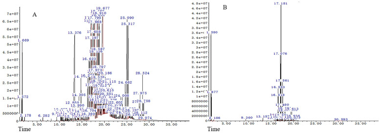 Figure 1
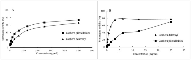 Figure 2
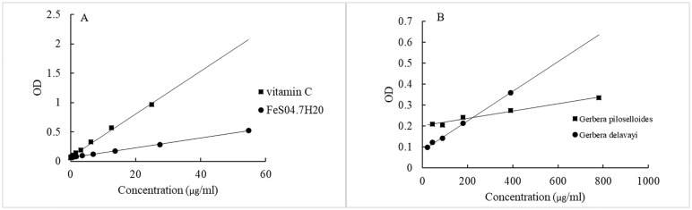 Figure 3
| No. | Components | Retention time (min) | RI | CAS number | Molecular formula |
|---|---|---|---|---|---|
| 1 | Ethanol | 1.58 | 61 | 64-17-5 | C2H6O |
| 2 | Ethyl ether | 1.677 | 73 | 60-29-7 | C4H10O |
| 3 | Ethyl acetate | 2.186 | 136 | 141-78-6 | C4H8O2 |
| 4 | (+)-alpha-Pinene | 9.36 | 1024 | 7785-70-8 | C10H16 |
| 5 | Adipic acid, di(trans-hex-3-enyl) ester | 13.157 | 1494 | No | C18H30O4 |
| 6 | Thymol | 15.686 | 1807 | 89-83-8 | C10H14O |
| 7 | 1-Ethyl-3-(propen-1-yl)adamantane | 16.244 | 1876 | No | C15H24 |
| 8 | (-)-Aristolene | 16.413 | 1897 | 6831-16-9 | C15H24 |
| 9 | (-)-alpha-Gurjunene | 16.518 | 1910 | 489-40-7 | C15H24 |
| 10 | Cycloisolongifolene | 16.583 | 1918 | No | C15H24 |
| 11 | cyclosativene | 16.874 | 1954 | 22469-52-9 | C15H24 |
| 12 | alpha-longipinene | 16.93 | 1961 | 5989-08-2 | C15H24 |
| 13 | 4-(2’, 4’, 4’-trimethyl-yciclo[4.1.0]hept-2’-en-3’-yl)-3-buten-2-one | 17.076 | 1979 | No | C14H20O |
| 14 | Berkheyaradulene | 17.181 | 1992 | 65372-78-3 | C15H24 |
| 15 | Cyperene | 17.351 | 2013 | 2387-78-2 | C15H24 |
| 16 | Longifolene | 17.48 | 2029 | 61262-67-7 | C15H24 |
| 17 | Caryophyllene | 17.561 | 2039 | 87-44-5 | C15H24 |
| 18 | Humulene | 18.021 | 2096 | 26259-79-0 | C15H24 |
| 19 | Carvacryl acetate | 18.15 | 2112 | 6380-28-5 | C12H16O2 |
| 20 | (+)-DELTA-CADINENE | 18.756 | 2187 | 483-76-1 | C15H24 |
| 21 | 2,3,5,6-tetramethyl-Phenol | 19.282 | 2252 | 527-35-5 | C10H14O |
| 22 | Neryl 2-methylbutanoate | 19.403 | 2267 | 51117-19-2 | C15H26O2 |
| 23 | Caryophyllenyl alcohol | 19.532 | 2283 | No | C15H26O |
| 24 | Caryophyllene oxide | 19.613 | 2293 | 1139-30-6 | C15H24O |
| 25 | humulene epoxide ii | 19.936 | 2333 | 19888-34-7 | C15H24O |
| 26 | Isocaryophillene | 20.073 | 2350 | 13877-93-5 | C15H24 |
| 1,1,1,3,5,5,5-Heptamethyltrisiloxane | |||||
| 27 | Total Identified | 30.593 | 3652 | 1873-88-7 | C7H22O2Si3 |
| Monoterpenes | 76.61 | ||||
| Sesquiterpenes | 0.31 | ||||
| Phenolic Compounds | 55.50 | ||||
| Aromatic Compounds | 2.73 | ||||
| Esters | 13.75 | ||||
| Other Compounds | 1.98 |
| No. | Components | Retention time (min) | RI | CAS number | Molecular formula |
|---|---|---|---|---|---|
| 1 | Ethanol | 1.572 | 60 | 64-17-5 | C2H6O |
| 2 | Ethyl ether | 1.669 | 72 | 60-29-7 | C4H10O |
| 3 | Ethyl Acetate | 2.178 | 135 | 141-78-6 | C4H8O2 |
| 4 | 2-Isopropoxyethanol | 6.282 | 643 | 109-59-1 | C5H12O2 |
| 5 | 3-Ethylidenecycloheptene | 9.36 | 1024 | – | C9H14 |
| 6 | Sabinene | 10.257 | 1135 | 3387-41-5 | C10H16 |
| 7 | 1-(4-Methylphenyl)ethanol | 10.354 | 1147 | 536-50-5 | C9H12O |
| 8 | 6-methyl-5-Hepten-2-one | 10.507 | 1166 | 110-93-0 | C8H14O |
| 9 | 2,2-dimethyl-3-octyne | 10.62 | 1180 | 19482-57-6 | C10H18 |
| 10 | Alpha -Phellandrene | 10.944 | 1220 | 99-83-2 | C10H16 |
| 11 | O-Cymene | 11.307 | 1265 | 527-84-4 | C10H14 |
| 12 | D-Limonene | 11.404 | 1277 | 5989-27-5 | C10H16 |
| 13 | Eucalyptol | 11.469 | 1285 | 470-82-6 | C10H18O |
| 14 | Beta-Ocimene | 11.719 | 1316 | 13877-91-3 | C10H16 |
| 15 | (Z)-linalool oxide (furanoid) | 12.196 | 1375 | 5989-33-3 | C10H18O2 |
| 16 | (+)-2-Carene | 12.713 | 1439 | – | C10H16 |
| 17 | cyclene | 12.85 | 1456 | 508-32-7 | C10H16 |
| 18 | N-methyl-2-pyrolidene | 13.376 | 1521 | 33838-11-8 | C5H9N |
| 19 | nerol oxide | 13.602 | 1549 | 1786-08-9 | C10H16O |
| 20 | 5-methyl-3-(1-methylethylidene)-1,4-Hexadiene | 13.99 | 1597 | – | C10H16 |
| 21 | (-)-Terpinen-4-ol | 14.086 | 1609 | 20126-76-5 | C10H18O |
| 22 | 3-methylene-1,5,5-trimethyl-cyclohexene | 14.256 | 1630 | 16609-28-2 | C10H16 |
| 23 | 2-hydroxy-4-methylbenzaldehyde | 14.305 | 1636 | 698-27-1 | C8H8O2 |
| 24 | 3-Carene | 14.773 | 1694 | 13466-78-9 | C10H16 |
| 25 | 2-isopropyl-4-methyl anisole | 14.927 | 1713 | 31574-44-4 | C11H16O |
| 26 | Citral | 15.347 | 1765 | 5392-40-5 | C10H16O |
| 27 | cis-Thujopsene | 15.735 | 1813 | 470-40-6 | C15H24 |
| 28 | 1-(4-Hydroxy-3-methylphenyl)ethanone | 15.969 | 1842 | 876-02-8 | C9H10O2 |
| 29 | 1,5-dimethyl-2,4-bis(1-methylethyl)-benzene | 16.276 | 1880 | 5186-68-5 | C14H22 |
| 30 | 1-Isopropyl-4,7-dimethyl-1,2,4a,5,8,8a-hexahydronaphthalene | 16.389 | 1894 | 5951-61-1 | C15H24 |
| 31 | (-)-Aristolene | 16.583 | 1918 | 6831-16-9 | C15H24 |
| 32 | Cedrene-V6 | 16.623 | 1923 | – | C15H24 |
| 33 | Aciphyllene | 16.704 | 1933 | – | C15H24 |
| 34 | cyclosativene | 17.011 | 1971 | 22469-52-9 | C15H24 |
| 35 | alfa.-Copaene | 17.116 | 1984 | 138874-68-7 | C15H24 |
| 36 | 4-(2’, 4’, 4’-trimethyl-yciclo[4.1.0]hept-2’-en-3’-yl)-3-buten-2-one | 17.197 | 1994 | – | C14H20O |
| 37 | Berkheyaradulene | 17.254 | 2001 | 65372-78-3 | C15H24 |
| 38 | (-)-Cyperene | 17.496 | 2031 | 2387-78-2 | C15H24 |
| 39 | Caryophyllene | 17.714 | 2058 | 87-44-5 | C15H24 |
| 40 | 1-(1,1-dimethylethyl)-4-ethyl-benzene | 17.795 | 2068 | 7364-19-4 | C12H18 |
| 41 | 2,4-diethyl-7,7-dimethylcyclohepta-1,3,5-triene | 17.868 | 2077 | – | C13H20 |
| 42 | 2,5-Dimethylchroman-4-one | 17.9 | 2081 | 69687-87-2 | C11H12O2 |
| 43 | beta-maaliene | 17.973 | 2090 | 489-29-2 | C15H24 |
| 44 | Humulene | 18.126 | 2109 | 6753-98-6 | C15H24 |
| 45 | Alloaromadendrene | 18.175 | 2115 | 25246-27-9 | C15H24 |
| 46 | rotundene | 18.207 | 2119 | – | C15H24 |
| 47 | beta-Panasinsene | 18.433 | 2147 | 56684-97-0 | C15H24 |
| 48 | Selina-3,7(11)-diene | 18.482 | 2153 | 6813-21-4 | C15H24 |
| 49 | gamma-selinene | 18.506 | 2156 | 515-17-3 | C15H24 |
| 50 | beta-Guaiene | 18.538 | 2160 | 88-84-6 | C15H24 |
| 51 | Alloaromadendrene | 18.587 | 2166 | 25246-27-9 | C15H24 |
| 52 | alpha.-Muurolene | 18.651 | 2174 | 31983-22-9 | C15H24 |
| 53 | (+)-Calarene | 18.797 | 2192 | 17334-55-3 | C15H24 |
| 54 | (+)-DELTA-CADINENE | 18.91 | 2206 | 483-76-1 | C15H24 |
| 55 | Guaia-9,11-diene | 18.974 | 2214 | – | C15H24 |
| 56 | Isolongifolene | 19.023 | 2220 | 1135-66-6 | C15H24 |
| 57 | (+)-α-murolene | 19.071 | 2226 | 17627-24-6 | C15H24 |
| 58 | 1, 1, 5-Trimethyl-1, 2-dihydronaphthalene | 19.128 | 2233 | – | C13H16 |
| 59 | 3-Methyl-2-butenoic acid, 4-methoxybenzyl ester | 19.241 | 2247 | – | C13H16O3 |
| 60 | (E)-3,7-Dimethylocta-2,6-dienyl ethyl carbonate | 19.338 | 2259 | – | C13H22O3 |
| 61 | [(2E)-3,7-dimethylocta-2,6-dienyl] butanoate | 19.629 | 2295 | 106-29-6 | C14H24O2 |
| 62 | Caparratriene | 19.694 | 2303 | – | C15H26 |
| 63 | 1,3-dimethyl-5-ethylbenzene | 19.782 | 2314 | 934-74-7 | C10H14 |
| 64 | .beta.-Guaiene | 19.895 | 2328 | 88-84-6 | C15H24 |
| 65 | Delta-Selinene | 19.968 | 2337 | 473-14-3 | C15H24 |
| 66 | Alloaromadendrene | 20.025 | 2344 | 025246-27-9 | C15H24 |
| 67 | Alpha-Elemene | 20.081 | 2351 | 5951-67-7 | C15H24 |
| 68 | dehydro-aromadendrene | 20.122 | 2356 | – | C15H22 |
| 69 | Xanthurenic acid | 20.186 | 2364 | 59-00-7 | C10H7NO4 |
| 70 | 4,8a-dimethyl-6-prop-1-en-2-yl-1,3,5,6,7,8-hexahydronaphthalen-2-one | 20.316 | 2380 | – | C15H22O |
| 71 | 1,2,3,5,6,7,8,8a-octahydro-1-methyl-6-methylene-4-(1methylethyl)naphthalene | 20.356 | 2385 | 150320-52-8 | C15H24 |
| 72 | 4-(2,3,4,6-Tetramethylphenyl)-3-buten-2-one | 20.429 | 2394 | – | C14H18O |
| 73 | Epizonarene | 20.501 | 2403 | 41702-63-0 | C15H24 |
| 74 | Longifolene | 20.566 | 2411 | 475-20-7 | C15H24 |
| 75 | Cedren-13-ol, 8- | 20.615 | 2417 | 18319-35-2 | C15H24O |
| 76 | 1,2,3a,6-Tetramethyloctahydrocyclopenta[c]pentalen-3(3ah)-one | 20.728 | 2431 | – | C15H24O |
| 77 | 7R,8R-8-Hydroxy-4-isopropylidene-7methylbicyclo[5.3.1]undec-1-ene | 20.946 | 2458 | – | C15H24O |
| 78 | 3,5,6,7,8,8a-hexahydro-4,8a-dimethyl-6-(1-methylethenyl)-2 Naphthalenone | 21.019 | 2467 | – | C15H22O |
| 79 | Longipinocarvone | 21.229 | 2493 | – | C15H22O |
| 80 | Cadina-1(10),6,8-triene | 21.326 | 2505 | 1460-96-4 | C15H22 |
| 81 | 2,2,7,7-tetramethyltricyclo[6.2.1.01,6]undec-5-en-4-one | 21.6 | 2539 | 23747-14-0 | C15H22O |
| 82 | 6-Isopropenyl-4,8a-dimethyl-1,2,3,5,6,7,8,8a-octahydronaphthalene-2,3-diol | 21.721 | 2554 | – | C15H24O2 |
| 83 | Corymbolone | 21.923 | 2579 | 97094-19-4 | C15H24O2 |
| 84 | 2,2,7,7-tetramethyltricyclo[6.2.1.01,6]undec-5-en-4-one | 22.02 | 2591 | 23747-14-0 | C15H22O |
| 85 | Fukinanolid | 22.247 | 2619 | 19906-72-0 | C15H22O2 |
| 86 | (2-hydroxy-5-methylphenyl)-(4-methoxyphenyl)methanone | 22.279 | 2623 | – | C15H14O3 |
| 87 | 6,10,14-trimethylpentadecan-2-one | 22.36 | 2633 | 502-69-2 | C18H36O |
| 88 | (2-Hydroxy-5-methylphenyl)(4-methoxyphenyl)methanone | 22.602 | 2663 | – | C15H14O3 |
| 89 | 1-Methyl-1-silolanyl heptanoate | 22.723 | 2678 | – | C12H24O2Si |
| 90 | 2,6-ditert-butylnaphthalene | 23.418 | 2764 | 3905-64-4 | C18H24 |
| 91 | n-Hexadecanoic acid | 23.644 | 2792 | 57-10-3 | C16H32O2 |
| 92 | N-Mesitytricyclo-[3.2.1.0(2.4)]octane-3-carboxamide | 24.008 | 2837 | 342394-51-8 | C18H23NO |
| 93 | Kaur-15-ene | 24.169 | 2857 | 5947-50-2 | C9H12N2O5S |
| 94 | 2-(4-methoxybenzoyl)-1,6-dimethyl-1,2,3,4-tetrahydropyrrolo[1,2-a]pyrazine | 24.202 | 2861 | – | C17H20N2O2 |
| 95 | Kaur-16-ene | 24.662 | 2918 | 562-28-7 | C20H32 |
| 96 | N-(1-adamantyl)-2-hydroxybenzamide | 25.09 | 2971 | 3728-06-1 | C17H21NO2 |
| 97 | 2,5-Diethylpyrazine | 25.317 | 2999 | 013238-84-1 | C8H12N2 |
| 98 | propoxy(dipropyl)phosphane | 25.454 | 3016 | 6418-60-6 | C9H21OP |
| 99 | o-Terphenyl | 25.535 | 3026 | 84-15-1 | C18H14 |
| 100 | N-1-Adamantyl-p-nitrobenzalimine | 25.987 | 3082 | – | C17H20N2O2 |
| 101 | 3-Adamantan-1-yl-3-oxo-propionitrile | 26.197 | 3108 | – | C13H14NO |
| 102 | Hexestrol | 26.294 | 3120 | 84-16-2 | C18H22O2 |
| 103 | 1-(4-Methoxyphenyl)-4,6-dimethyl-2(1H)-pyrimidinone | 27.773 | 3303 | 74360-11-5 | C13H14N2O2 |
| 104 | 6,6-Diphenylfulvene | 27.975 | 3328 | 2175-90-8 | C18H14 |
| 105 | Triphenylene | 28.112 | 3345 | 217-59-4 | C18H12 |
| 106 | Benz[a]anthracene | 28.524 | 3396 | 56-55-3 | C18H12 |
| 107 | dimethylphenylsilane | 28.758 | 3425 | 766-77-8 | C8H11Si |
| 108 | 2,6-dimethylocta-2,4,6-triene | 29.074 | 3464 | 673-84-7 | C10H16 |
| Total identified | 95.06 |
| Compound | Content (%) | |
|---|---|---|
|
|
| |
| (-)-Aristolene | 0.357 | 1.611 |
| cyclosativene | 0.753 | 7.831 |
| 4-(2’, 4’, 4’-trimethyl-yciclo[4.1.0]hept-2’-en-3’-yl)-3-buten-2-one | 12.862 | 1.715 |
| Berkheyaradulene | 32.025 | 1.224 |
| Cyperene | 0.701 | 9.700 |
| Naphthalene, 1,2,3,5,6,8a-hexahydro-4,7-dimethyl-1-(1-methylethyl)-, (1S-cis)- | 0.268 | 3.654 |
| Humulene | 1.761 | 1.284 |
| Caryophyllene | 6.782 | 0.77 |
| Total (%) | 55.509 | 27.789 |
| Sample | TPC (mg eq gallic acid/mL oil) | FRAP (mg eq vitaminC/mL oil) | IC50 values | |
|---|---|---|---|---|
| DPPH (mg/mL) | ABTS (μg/mL) | |||
|
| 27.4 ± 2* | 7.8 ± 1* | 69.5 ± 1*** | 81 ± 5* |
|
| 68.6 ± 4* | 19.7 ± 5* | 0.7 ± 0.002*** | 105.8 ± 11** |
| Vitamin C | N.T. | N.T. | 0.003 ± 0.0001*** | 1.5 ± 0.02*** |
| Microbial Strains | MIC (mg/mL) for EO from | MIC (mg/mL) for EO from | MIC (mg/mL) for Chloramphenicol |
|---|---|---|---|
|
| 12.5 | 12.5 | 5 |
|
| 6.3 | 6.3 | 10 |
|
| 12.5 | 12.5 | 5 |
|
| 6.3 | 12.5 | 5 |
|
| 12.5 | 12.5 | 10 |
Peer Reviews
No public reviews on file for this paper yet. If you reviewed it on a platform where reviews are public (OpenReview, ICLR, NeurIPS, ICML), you can paste yours below so the community can read it here.
Videos
No videos yet. Explain this paper in a talk, walkthrough, or lecture? Add one.
Taxonomy
TopicsEssential Oils and Antimicrobial Activity · Ethnobotanical and Medicinal Plants Studies · Sesquiterpenes and Asteraceae Studies
Introduction
1
The relentless progression of antibiotic resistance, particularly among multidrug-resistant bacterial strains, poses a significant threat to global health, underscoring the urgent need for the continuous development and discovery of new antimicrobial materials (Baran et al., 2023). While the “Antibiotic Era” may be waning, the potential of medicinal plants as a source for novel antimicrobial agents remains a beacon of hope. Through the intricate process of photosynthesis, plants synthesize a wealth of organic matter and secondary metabolites, which exhibit a broad spectrum of biological activities. These compounds lay the pharmacological groundwork for the prevention and treatment of diseases (Petric et al., 2020; Rehman et al., 2020). With many medicinal plants recognized for their safety, efficacy, and minimal side effects, the exploration of their bioactive compounds for antimicrobial properties is not only imperative but also a promising avenue in the fight against multidrug-resistant bacteria (Bouarab Chibane et al., 2019).
Essential oils (EOs), a type of secondary metabolite produced by aromatic plants, exhibit a spectrum of biological activities, including antibacterial, antioxidant, anti-inflammatory, enzyme inhibitory, sedative, anxiolytic, and antidepressant properties (Zengin et al., 2019; Liu Y. et al., 2024). EOs are utilized as natural remedies for the treatment of infectious diseases and as flavoring agents in food, offering a green and healthy alternative (Coelho et al., 2023; Wu et al., 2024). Due to their remarkable biological activities, EOs from medicinal plants are of great interest to scientists seeking to identify new phytochemical bioactive molecules that align with biodiversity and medicinal needs (Oliveira de Veras et al., 2020).
Gerbera Cass., a member of the Compositae family (Mutisieae Cass.), comprises approximately 80 species ranging from Africa to East Asia, with 20 species found in China, predominantly in the southwestern region (Zhao et al., 2024). Gerbera piloselloides (L.) Cass. and Gerbera delavayi Franch. are perennial herbs within the Gerbera genus. G. piloselloides is known for its heat-clearing, detoxifying, cough-relieving, phlegm-resolving, and circulation-regulating properties (Zhao et al., 2022; Liu C. et al., 2024). Traditionally, it is used in southwestern China to treat cough and sore throat when mixed with honey and also serves as a flavoring agent in winemaking and meat cooking due to its pleasant aroma (Zhou et al., 2022). The plant’s bioactive compounds, including caffeic acid derivatives, parasorboside derivatives, coumarins, and flavonoids, have been isolated through activity-guided isolation (Wang et al., 2014). The EO of G. piloselloides, EOgp, has been analyzed by GC-MS and found to contain fatty acids, terpenes, and aromatic compounds (Tang et al., 2003).
G. delavayi, found in open areas and forest margins at altitudes of 1800 to 3200 meters, was historically known as “ignited flowers” or “fireweed” due to its leaf’s combustion-supporting properties (Xu et al., 2017). The soft fiber on the back of its leaves is used in hand-weaving (Zheng et al., 2017). Beyond its use in spinning, G. delavayi holds significance in medicine and ornamental purposes. Gerbera species in China are noted for their antitussive, antipyretic, hemostatic, circulatory, and anti-inflammatory effects (Wu et al., 2005). The ethanol extract of G. delavayi has led to the isolation of two new coumarin compounds, gerdelavins A and B, along with 13 known compounds (Liu et al., 2010). Coumarins, characterized by their benzopyrone core, interact with various enzymes and receptors in organisms through weak bonds, conferring a broad range of medicinal potential, including antibacterial, antitumor, and anticoagulant activities (Balewski et al., 2021; Citarella et al., 2024).
In this context, our study endeavors to delve deeper into the properties of two lesser-studied Gerbera species, G. piloselloides and G. delavayi. The objective was to assess the EO compositions and to explore their antioxidant and antimicrobial potential. Notably, there is a paucity of literature documenting the biological activities of the EOs from these two plant species. Consequently, this investigation stands as the first comprehensive examination of the biological activities of the extracted EOs from G. piloselloides and G. delavayi, marking a significant contribution to the existing knowledge base.
Materials and methods
2
Plant material
2.1
To obtain a comprehensive representation of the chemical profile, the entire plants of G. piloselloides and G. delavayi, encompassing leaves, stems, roots, and rhizomes, were collected from the Stone Forest region of China. Plant materials from two Gerbera species, were meticulously collected in the Stone Forest region of China. The sampling locations were at elevations of 2316 m (24°81′10.55″ N, 103°30′12.83″ E) for G. piloselloides and 1689 m (24°46′27.55″ N, 103°17′18.83″ E) for G. delavayi, within Shilin County, Kunming, Yunnan Province, in July 2019. The taxonomic identification of these species was conducted by the Professor Huifeng Sun from Heilongjiang University of Chinese Medicine in Harbin, China.
For posterity and to facilitate future studies, voucher specimens were meticulously archived in the Herbarium of the College of Veterinary Medicine. The voucher numbers assigned to G. piloselloides and G. delavayi are 2031 and 2032, respectively. Following collection, the herbal materials were subjected to natural drying at room temperature. Subsequently, they were finely pulverized using a grinder and preserved at a refrigerated temperature of 4 °C, awaiting subsequent utilization in experimental procedures.
Extraction of essential oils
2.2
The essential oils (EOs) from G. piloselloides and G. delavayi were extracted using the hydrodistillation method, as described by Semerdjieva et al. (2019). For the extraction process, a precise amount of 100 grams of dried plant material was combined with 1000 mL of distilled water in a flask. The extraction was conducted for a duration of 8 h, commencing once the water reached boiling point. Following extraction, the EOs were separated from the aqueous phase with ethyl ether, dried over anhydrous sodium sulfate, filtered, and then subjected to evaporation of the ethyl ether in an oven at 40 °C for one hour. The resulting EO was transferred to amber vials and stored at -20 °C. The yield percentage (w/w) of the oil was determined based on the initial weight of the plant material used.
GC/MS analysis
2.3
The compositional analysis of the EOs was performed using an Agilent Technologies Gas Chromatograph model 7697A, equipped with a triple quadrupole detection system and a split-splitless injection port. The chromatographic separation was achieved on a HP-5MS fused silica capillary column (30 m × 250 μm × 0.25 μm) coupled with an Agilent MS Detector. The column temperature program began at 40 °C for 5 min, followed by an increase to 280 °C at a rate of 10 °C/min. An injection volume of 0.8 μL was used with a split ratio of 1: 20, and helium was employed as the carrier gas at a constant flow rate of 20 mL/min. Mass spectra were acquired at an electron energy of 70 eV, with the ion source temperature set at 250 °C. The mass spectra data were recorded within the mass-to-charge ratio (m/z) range of 44-550.
The identification of the EO compounds was accomplished by comparing their retention times and mass spectra with reference data in the NIST mass spectra library. The relative percentage contents of the individual compounds were quantified based on the peak areas in the GC-MS chromatograms, following the methodology described by Thabet et al. (2022).
The identification of the EO compounds was accomplished by comparing their retention times and mass spectra with reference data in the NIST mass spectra library. The retention indices were calculated using the linear retention index method, with a mixture of n-alkanes (C8-C20) at a concentration of 1 μg/mL as the reference compounds. The relative percentage contents of the individual compounds were quantified based on the peak areas in the GC-MS chromatograms, following the methodology described by Thabet et al. (2022).
Estimation of total polyphenolic content
2.4
The TPC was quantified using the Folin-Ciocalteu method adapted for a 96-well microplate format (Larrazabal-Fuentes et al., 2019). Initially, the EO sample (500 μg/mL) was combined with 10% (v/v) Folin-Ciocalteu reagent at a ratio of 1: 5 and allowed to stand for 5 min. Subsequently, a sodium carbonate solution was added to the mixture at a volume four times that of the sample and the mixture was shaken for 1 min. After incubation for 1 h at 25 °C, the absorbance was recorded at 765 nm using a microplate reader. A calibration curve was generated using gallic acid dilutions ranging from 0 to 1000 μg/mL. The results were expressed as milligrams of gallic acid equivalents per milliliter of EO.
Evaluation of antioxidant activities
2.5
The antioxidant potential of the EOs was assessed using the ferric reducing antioxidant power (FRAP) assay, 2,2’-azinobis-(3-ethylbenzothiazoline-6-sulfonic acid) (ABTS) assay, and 1,1-diphenyl-2-picrylhydrazyl (DPPH) scavenging assay, in conjunction with the determination of the TPC. Vitamin C was employed as the standard reference. The protocols outlined below were adapted for use with 96-well microplates. All assays were conducted in triplicate.
DPPH radical scavenging activity assay
2.5.1
The DPPH radical scavenging capacity of the EOs was evaluated using the methodology of Larrazabal-Fuentes et al. (2019). The percent inhibition (I%) was calculated with the formula: I% = [(Ac - As)/(Ac)] × 100, where Ac is the absorbance of the control and As is the absorbance of the sample. The results were reported as IC_50_ values, representing the concentration of EO (μg/mL) required to inhibit 50% of the DPPH radicals in the solution, determined through linear regression analysis of the percentage of residual DPPH versus sample concentration.
FRAP assay
2.5.2
The FRAP assay was performed as described by Tian et al. (2019). An extract solution (500 μg/mL, 10 μL) was mixed with freshly prepared FRAP solution (70 μL), and the change in absorbance was measured at 593 nm after a 30-min incubation at 37 °C. Standard solutions of FeSO_4_·7H_2_O (0-500 μg/mL) and vitamin C (0-200 μg/mL) were used to construct the calibration curve. FRAP results were expressed as milligrams of vitamin C equivalent per milliliter of EO.
ABTS scavenging activity
2.5.3
The ABTS scavenging activity of the EO was determined following the procedures of Kıvrak (2014). ABTS radical cation (ABTS+) was generated by reacting ABTS (7 mM) with potassium persulfate (2.45 mM) at room temperature in the dark for 16 h. The ABTS+ solution was then diluted with ethanol to achieve an absorbance of 0.700 ± 0.005 at 734 nm. This solution (160 μL) was mixed with 40 μL of EO (0-20000 μg/mL), and the absorbance was measured at 734 nm after a 30-minute incubation at 30 °C. Vitamin C at various concentrations (0-200 μg/mL) served as the reference. The percentage scavenging of ABTS radicals was calculated using the equation from the DPPH assay. The results were expressed as IC_50_ values, calculated based on the linear regression of the percentage of residual ABTS versus sample concentration.
Evaluation of antibacterial activity
2.6
Microbial strains employed
2.6.1
The antimicrobial efficacy of the EOs was assessed against a panel of bacterial strains, including S. aureus CMCC26003, Listeria ATCC 19111, Salmonella CVCC541, E. coli CVCC10141, and P. multocida C48-1. These strains were obtained from the Harbin Institute of Veterinary Medicine (Harbin, Heilongjiang, China).
Determination of minimal inhibitory concentrations
2.6.2
The MICs of the EOs were determined using the broth microdilution method, as outlined in the CLSI protocols M60 (CLSI, 2017) and M100 (CLSI, 2018). The procedure involved preparing a stock solution of EO at 25 mg/mL in a mixture of 20% dimethyl sulfoxide (DMSO) and 80% distilled water. Initially, 100 µL of this stock solution was added to the first well of a 96-well plate, followed by serial two-fold dilutions to achieve concentrations ranging from 25 to 0.05 mg/mL (Cui et al., 2018). Subsequently, each bacterial strain was inoculated into LB broth to achieve a McFarland standard of 0.5, diluted 100-fold, and then added to the wells at a volume of 100 µL per well. The MIC was defined as the lowest concentration of EO that inhibited visible growth of the bacterial strains after an incubation period of 16–18 h at 37 °C. Chloramphenicol, at concentrations ranging from 10 to 0.04 mg/mL, was used as a positive control, while a solution of 20% DMSO-80% distilled water served as the negative control. All experiments were conducted in triplicate to ensure accuracy and reproducibility.
Statistical analysis
2.7
Significance was determined at a p-value threshold of < 0.05. Data were processed using GraphPad Prism^®^ version 7.0 and presented as mean ± standard deviation (SD). To ascertain statistically significant differences among the groups, a one-way analysis of variance (ANOVA) was performed.
Results and discussion
3
Chemical composition of essential oils
3.1
The medicinal parts of two Gerbera species, namely the whole plants of G. piloselloides and G. delavayi, were subjected to hydrodistillation, yielding yellow EOs with distinctive odors. The yields for G. piloselloides and G. delavayi were 0.14% (w/w) and 0.26% (w/w), respectively. The chemical compositions of these EOs were elucidated using GC/MS. The compositional percentages of the EOs from G. piloselloides and G. delavayi are presented in Tables 1 and 2, respectively. The total ion chromatograms for the EOs of both species (EOgp and EOgd) are depicted in Figure 1. GC/MS analysis of EOgp identified 24 components, with berkheyaradulene (32.03%), 4-(2’, 4’, 4’-trimethyl-cyclo[4.1.0]hept-2’-en-3’-yl)-3-buten-2-one (12.86%), caryophyllene (6.78%), and cycloisolongifolene (5.30%) as the principal constituents. In contrast, EOgd comprised 100 components, with butanoic acid, 3,7-dimethyl-2,6-octadienyl ester, (E)- (10.50%), cyperene (9.70%), β-panasinsene (7.13%), benzamide, N-(1-adamantyl)-2-hydroxy- (6.12%), and benzene, 1-(1,1-dimethylethyl)-4-ethyl- (5.31%) as the predominant compounds.
GC/MS profiles of the essential oils from Gerbera piloselloides (A) and Gerbera delavayi (B).
In a prior phytochemical study, the researchers documented the presence of 17 volatile organic components in G. piloselloides, encompassing fatty acids, terpenes, and aromatic compounds. Notably, neryl (S) -2-methylbutanoate (35.99%), 4-hydroxy-3-methylacetophenone (8.74%), and n-hexadecanoic acid (7.48%) emerged as the predominant constituents of the plant’s essential oil (Luo et al., 2013). Previous studies have identified various volatile organic compounds in specific parts of G. piloselloides, such as leaves, caudices, and roots (Tang et al., 2003). However, our study focused on the essential oils extracted from the whole plants of G. piloselloides and G. delavayi. The observed discrepancies in the identified compounds may be attributed to the varying environmental conditions of the plant collection sites and the inclusion of multiple plant parts in our analysis.
Previous research on Gerbera has revealed the presence of coumarins, sesquiterpenoids, triterpenoids, and cyanogenic glycosides (Liu et al., 2010). Despite the distinct EO profiles of the two Gerbera species, eight compounds, including (-)-aristolene, 4-(2’, 4’, 4’-trimethyl-cyclo[4.1.0]hept-2’-en-3’-yl)-3-buten-2-one, berkheyaradulene, cyclosativene, cyperene, naphthalene, 1,2,3,5,6,8a-hexahydro-4,7-dimethyl-1-(1-methylethyl)-, (1S-cis)-, humulene, and caryophyllene, are common to both, as detailed in Table 3.
Berkheyaradulene is particularly abundant in G. piloselloides (10.50%), representing a sesquiterpene hydrocarbon with an unusual carbon skeleton characterized by a bridgehead carbon connected to three rings, also found in other Asteraceae plants (Szöke et al., 2004). Caryophyllene, notable for its cyclobutane ring, a rare occurrence in nature, is often accompanied by isocaryophyllene and α-humulene, its ring-opened isomer (Taherpour et al., 2010). Cyperene, a tetracyclic sesquiterpene, possesses unique properties such as sterilizing, antioxidant, anticarcinogenic, and immune-boosting functions (Skała et al., 2016; Hu et al., 2017). Thymol, with its thyme oil-like aroma, may contribute to the use of G. piloselloides in winemaking and meat cooking. Thymol’s expectorant properties have been documented, and it also exhibits bactericidal effects, suggesting its potential in treating bronchitis and whooping cough (Zhou et al., 2019). Furthermore, thymol holds promise for applications in the preservatives industry, as an insect repellent, and in the perfume industry (Roufegarinejad et al., 2018; Reyhani et al., 2022; Dadé et al., 2023).
Antioxidant capacity of essential oils
3.2
EOs are integral aromatic constituents found in herbs and spices, conferring them with a range of biological activities, including antimicrobial, antifungal, antioxidant, and anti-inflammatory effects (Valdivieso-Ugarte et al., 2019). However, the composition of EOs in these herbs is intricate, lacking a straightforward and precise method for a comprehensive and objective evaluation of the antioxidant capacity of traditional Chinese medicines. Consequently, a variety of antioxidant assays are necessary to profile the total antioxidant potential of natural extracts in this context.
We assessed the antioxidant potential of EOgp and EOgd by evaluating their efficacy in scavenging the stable free radicals ABTS and DPPH. The radical scavenging activities of the EOs are depicted in Figures 2A and 2B, respectively. The concentrations of the EOs required to inhibit each radical by 50% (IC_50_) are presented in Table 4. Notably, G. delavayi exhibited a significantly higher DPPH free radical scavenging ability (IC_50_ 0.7 mg/mL) compared to G. piloselloides (IC_50_ 69.5 mg/mL). However, both G. piloselloides and G. delavayi demonstrated similar ABTS free radical scavenging activity, with IC_50_ values of 81 µg/mL and 105.8 µg/mL, respectively. It is documented that certain compounds with ABTS scavenging capability may not exhibit DPPH scavenging activity, which could account for the observed results (Borah et al., 2019).
Radical scavenging activity of the essential oils from Gerbera piloselloides and Gerbera delavayi against the ABTS radical (A) and DPPH (B). Data are represented as mean standard deviation (SD) of triplicate experiments.
The reducing capability of the extracts was determined using a microplate reader to track the conversion of Fe^3+^ to Fe^2+^ in the presence of the extracts. An increase in absorbance is indicative of the extract’s reducing power. The influence of antioxidant concentration on FRAP inhibition is summarized in Figure 3. FRAP results were expressed as milligrams of vitamin C equivalent per milliliter of oil, and TPC data are provided in Table 4. G. delavayi displayed a superior antioxidant capacity, with 19.7 mg eq vitamin C/mL oil, compared to G. piloselloides (7.8 mg eq vitamin C/mL oil). The FRAP antioxidant activities were directly proportional to the TPC, with G. piloselloides and G. delavayi exhibiting TPC values of 27.4 mg eq gallic acid/mL oil and 68.6 mg eq gallic acid/mL oil, respectively. Studies by other researchers have also linked the antioxidant activity of Solanum elaeagnifolium to its TPC (Bouslamti et al., 2022). These findings suggest that TPC compounds contribute significantly to the antioxidant activity of G. delavayi.
Concentration-dependent effects of antioxidants on the inhibition of the FRAP assay. (A) shows the correlation coefficients (r²) for vitamin C (r² = 0.996) and FeSO4·7H2O (r² = 0.999); (B) depicts the correlation coefficients for Gerbera piloselloides (r² = 0.978) and FeSO4·7H2O (r² = 0.998).
Antimicrobial activity of essential oils
3.3
Bacterial infections continue to be a leading cause of mortality worldwide, a situation exacerbated by the persistent emergence of antibiotic resistance (Huemer et al., 2020). EO components derived from medicinal plants are noted for their high biological activity, and the quest for alternative antimicrobial agents to replace antibiotics has become a focal point of contemporary research (Coimbra et al., 2022).
This study assessed the antimicrobial potential of G. piloselloides and G. delavayi by evaluating their inhibitory effects against Listeria, S. aureus, Salmonella, P. multocida, and E. coli. The minimum inhibitory concentrations (MICs) of the EOs against these microbial strains are detailed in Table 5. The data reveal that EOgp demonstrated inhibitory activity against Listeria ATCC 19111, S. aureus CMCC26003, Salmonella CVCC541, P. multocida C48-1, and E. coli CVCC10141 with MICs of 6.3 mg/mL, 12.5 mg/mL, 12.5 mg/mL, 6.3 mg/mL, and 12.5 mg/mL, respectively. Similarly, Eogd exhibited efficacy against the same pathogens with MIC values of 6.3 mg/mL, 12.5 mg/mL, 12.5 mg/mL, 6.3 mg/mL, and 6.3 mg/mL, respectively. Notably, the antimicrobial potency of both EOs against Listeria surpassed that of chloramphenicol.
The biological effects of EOs are a consequence of the synergistic interaction of all molecules within the oil, and it is erroneous to attribute these effects to a single compound (Melo et al., 2020). The predominant components identified in both plant EOs were terpenes, natural products that serve diverse roles in various organisms and exhibit a wide array of structural diversity. Listeria has long been implicated as a primary agent of foodborne diseases in humans and animals. Cho et al. (2020) reported on the combined activities of gaseous oregano and thyme thymol EOs against Listeria monocytogenes. In the study by Said et al. (2016), oxygenated terpenes such as chamazulene-a degradation product, β-thujone, and camphor were identified as the main components of bioactive oils with antibacterial activity against Listeria monocytogenes. In the present manuscript, we also observed a high terpenoid content in both EOgp and EOgd, which may be responsible for their significant inhibitory effects against Listeria. The presence of these oxygenated terpenes in our extracts aligns with the findings of Said et al. (2016), suggesting that these compounds could be key contributors to the antibacterial properties observed. Our data further support the potential of natural plant-derived EOs as agents for controlling Listeria monocytogenes in antibacterial applications. However, it is important to note that while these EOs show promise, their safety profile must be thoroughly investigated before they can be considered for practical use.
Conclusion
4
In the present investigation, we assessed the chemical constituents, as well as the antioxidant and antimicrobial properties, of EOs extracted from the whole plants of G. piloselloides (EOgp) and G. delavayi (EOgd) via hydrodistillation. Our findings reveal that both EOgd and EOgp exhibit significant antioxidant capabilities and demonstrate differential inhibitory effects against five tested microbial strains. Notably, both essential oils exerted potent antibacterial effects against Listeria monocytogenes in vitro. These findings contribute to the growing body of evidence supporting the potential of these species as natural sources for the development of therapeutic products. Further research is needed to explore the specific mechanisms of action and safety profiles of these essential oils.
The reference list from the paper itself. Each links out to its DOI / PubMed record.
- 1BalewskiŁ.Szulta S.Jalińska A.Kornicka A. (2021). Recent advances in coumarin-metal complexes with biological properties. Front. Chem. 9. doi: 10.3389/fchem.2021.781779 PMC 867181634926402 · doi ↗ · pubmed ↗
- 2Baran A.Kwiatkowska A.Potocki L. (2023). Antibiotics and bacterial resistance-A short story of an endless arms race. Int. J. Mol. Sci. 24, 5777. doi: 10.3390/ijms 24065777 36982857 PMC 10056106 · doi ↗ · pubmed ↗
- 3Borah A.Paw M.Gogoi R.Loying R.Sarma N.Munda S.. (2019). Chemical composition, antioxidant, anti-inflammatory, anti-microbial and in-vitro cytotoxic efficacy of essential oil of Curcuma caesia Roxb. leaves: An endangered medicinal plant of North East India. Ind. Crops Prod. 129, 448–454. doi: 10.1016/j.indcrop.2018.12.035 · doi ↗
- 4Bouarab Chibane L.Degraeve P.Ferhout H.Bouajila J.Oulahal N. (2019). Plant antimicrobial polyphenols as potential natural food preservatives. J. Sci. Food Agric. 99, 1457–1474. doi: 10.1002/jsfa.9357 30206947 · doi ↗ · pubmed ↗
- 5Bouslamti M.El Barnossi A.Kara M.Alotaibi B. S.Al Kamaly O.Assouguem A.. (2022). Total polyphenols content, antioxidant and antimicrobial activities of leaves of Solanum elaeagnifolium Cav. from Morocco. Molecules 27, 4322. doi: 10.3390/molecules 27134322 35807566 PMC 9268098 · doi ↗ · pubmed ↗
- 6Cho Y.Kim H.Beuchat L. R.Ryu J. H. (2020). Synergistic activities of gaseous oregano and thyme thymol essential oils against Listeria monocytogenes on surfaces of a laboratory medium and radish sprouts. Food Microbiol. 86, 103357. doi: 10.1016/j.fm.2019.103357 31703857 · doi ↗ · pubmed ↗
- 7Citarella A.Vittorio S.Dank C.Ielo L. (2024). Syntheses, reactivity, and biological applications of coumarins. Front. Chem. 12. doi: 10.3389/fchem.2024.1362992 PMC 1090986138440776 · doi ↗ · pubmed ↗
- 8CLSI (2017). “Performance standards for antifungal susceptibility testing of yeasts,” in CLSI Supplement M 60, 1 th (Clinical Laboratory Standards Institute, Wayne, PA).