Detecting Breast Cancer via Innovative Magnetic Resonance Elastography with External Vibrations to the Back
Emi Yamaga, Tomoyuki Fujioka, Leona Katsuta, Makiko Hayashi, Katsura Yamamuro, Yuichi Kumaki, Kumiko Hayashi, Goshi Oda, Kazunori Kubota, Ukihide Tateishi

Abstract
Genes, proteins, chemicals, diseases, species, mutations and cell lines named across the full text — each resolved to its canonical identifier and authoritative record.
Click any figure to enlarge with its caption.
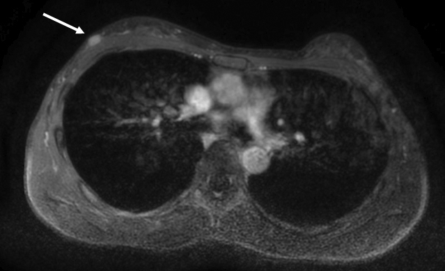 Figure 1
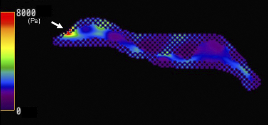 Figure 2
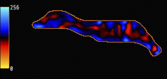 Figure 3
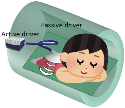 Figure 4
Peer Reviews
No public reviews on file for this paper yet. If you reviewed it on a platform where reviews are public (OpenReview, ICLR, NeurIPS, ICML), you can paste yours below so the community can read it here.
Videos
No videos yet. Explain this paper in a talk, walkthrough, or lecture? Add one.
Taxonomy
TopicsUltrasound Imaging and Elastography · Infrared Thermography in Medicine · Digital Imaging for Blood Diseases
A woman in her 40s, with a mass detected in her right breast through screening ultrasound, underwent breast magnetic resonance elastography (MRE); the results revealed a hard, elastic mass, which was later confirmed as invasive ductal carcinoma (Figure 1, 2 and 3). The patient received hormone therapy after surgery and remains relapse-free since then. Elastography measures tissue stiffness by assessing the propagation of external vibrations and is implemented using ultrasound and magnetic resonance imaging ^(1)^. Although MRE is clinically used to assess liver cirrhosis, its application for breast imaging remains unclear ^(2)^. Previous reports required specialized devices to apply external vibrations directly to the breast for performing breast MRE ^(3), (4)^. However, we have developed a simplified approach that enables breast MRE by applying external vibrations from the back using a passive driver originally designed for liver applications (Figure 4). This method considerably contributed to a highly confident diagnosis of breast cancer.
Article Information
Conflicts of Interest
None
Sources of Funding
This work was supported by JSPS KAKENHI grant number JP21K15842.
Acknowledgement
We used GPT-4 (https://chat.openai.com/) for Japanese to English translation and English proofreading. The generated text was reviewed, revised, and proofread by the authors. We would like to express our gratitude to “Irasutoya” for allowing us to use their illustrations in this paper.
Author Contributions
All the authors cared for the patient, as well as wrote and approved the final manuscript.
Approval by Institutional Review Board (IRB)
The Ethics Review Committee of the Faculty of Medicine, Institute of Science Tokyo approved this study (approval number: M2020-206). All procedures performed involving the patient were in accordance with the ethical standards of the institutional and/or National Research Committee and with the 1964 Declaration of Helsinki and its later amendments or comparable ethical standards.
Informed Consent
Informed consent was obtained from the patient.
The reference list from the paper itself. Each links out to its DOI / PubMed record.
- 1Muthupillai R, Lomas DJ, Rossman PJ, et al. Magnetic resonance elastography by direct visualization of propagating acoustic strain waves. Science. 1995;269(5232):1854-7.7569924 10.1126/science.7569924 · doi ↗ · pubmed ↗
- 2Moura Cunha G, Fan B, Navin PJ, et al. Interpretation, reporting, and clinical applications of liver MR elastography. Radiology. 2024;310(3):e 231220.38470236 10.1148/radiol.231220 PMC 10982829 · doi ↗ · pubmed ↗
- 3Bohte AE, Nelissen JL, Runge JH, et al. Breast magnetic resonance elastography: a review of clinical work and future perspectives. NMR Biomed. 2018;31(10):e 3932.29846986 10.1002/nbm.3932 · doi ↗ · pubmed ↗
- 4Kim HJ, Kim HH, Choi WJ, et al. Correlation of shear-wave elastography parameters with the molecular subtype and axillary lymph node status in breast cancer. Clin Imaging. 2023;101:190-9.37418896 10.1016/j.clinimag.2023.06.006 · doi ↗ · pubmed ↗