Lack of serologic evidence of orthoflavivirus infection in dogs with meningoencephalitis of unknown origin and steroid-responsive meningitis-arteritis in the Netherlands
Koen M. Santifort, Kiki Streng, Niklas Bergknut, Iris Van Soens, Marta Plonek, Wim H. M. van der Poel

TL;DR
This study found no evidence of orthoflavivirus infection in dogs with certain neurological conditions in the Netherlands.
Contribution
The study provides new serologic data ruling out orthoflavivirus as a cause of specific neurological diseases in Dutch dogs.
Findings
Serum samples from 12 dogs with MUO tested negative for orthoflaviviruses.
No evidence of West Nile, Usutu, or tick-borne encephalitis viruses was found.
Results suggest orthoflavivirus is not a cause of MUO or SRMA in the Netherlands.
Abstract
The pathogenesis of meningoencephalomyelitis of unknown origin (MUO) and steroid-responsive meningitis-arteritis (SRMA) in dogs remains enigmatic. Numerous studies have attempted and failed to identify (viral) pathogens in samples from MUO- or SRMA-diagnosed dogs. Orthoflavivirus-associated meningoencephalitis or meningoencephalomyelitis has been diagnosed in dogs in several European countries. We investigated serologic evidence for orthoflavivirus infection in dogs with clinical diagnoses of MUO or SRMA in the Netherlands. Twelve dogs with a clinical diagnosis of MUO based on signalment, neurologic examination, MRI studies, CSF analysis, and response to treatment were included in the study (age range: 1–11 y; 4 females, 8 males; weight range: 8–44 kg). Serum samples from all 12 dogs tested negative in a commercial competitive ELISA and virus neutralization tests for West Nile virus,…
Genes, proteins, chemicals, diseases, species, mutations and cell lines named across the full text — each resolved to its canonical identifier and authoritative record.
Click any figure to enlarge with its caption.
 Figure 1
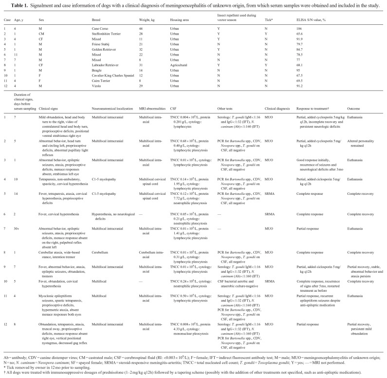 Figure 2
- —Toegepaste en Technische Wetenschappen, NWOhttps://doi.org/10.13039/501100024872
Peer Reviews
No public reviews on file for this paper yet. If you reviewed it on a platform where reviews are public (OpenReview, ICLR, NeurIPS, ICML), you can paste yours below so the community can read it here.
Videos
No videos yet. Explain this paper in a talk, walkthrough, or lecture? Add one.
Taxonomy
TopicsHerpesvirus Infections and Treatments · Viral Infections and Vectors · Rabies epidemiology and control
The terms meningoencephalomyelitis of unknown origin (MUO) and steroid-responsive meningitis-arteritis (SRMA) refer to inflammatory disorders of the CNS. MUO variants are characterized and classified histologically as necrotizing meningoencephalitis, necrotizing leukoencephalitis, and granulomatous meningoencephalitis.^ 8 ^ MUO can account for ~50% of CNS inflammatory disorders in dogs.^ 11 ^ To date, it is deemed most likely that MUO and SRMA are autoimmune disorders.^2,3,8^ Indeed, autoantibodies against neuronal and glial antigens have been identified in some cases.^2,3^ Nevertheless, it is possible that, in some patients, MUO has an infectious cause that is not identified by clinical or histologic tests. Orthoflaviviruses such as tick-borne encephalitis virus (TBEV; Flaviviridae, Orthoflavivirus encephalitidis) and West Nile virus (WNV; Flaviviridae, Orthoflavivirus nilense) have caused meningoencephalitis in human patients in the Netherlands since ~2015.^19,23^ Recognizing the emerging relevance of orthoflaviviruses in clinical veterinary medicine,^9,10,12,13,15,16,18,20,21^ we investigated serologic evidence for orthoflavivirus infection in dogs with MUO in the Netherlands.
Sampling of the animals in our study was performed under legislation (license AVD40100202114384) of the Dutch Central Authority for Scientific Procedures on Animals (CCD), and the experimental plan was approved by the Animal Welfare Body of Wageningen University and Research prior to the start of the sampling. For our prospective study, we included dogs with a clinical diagnosis of MUO or SRMA based on signalment, neurologic examination, and either or both MRI studies, and CSF analysis between May 2021 and May 2022. This subset of dogs was part of a larger serosurvey set up to investigate the seroprevalence of orthoflaviviruses in horses and dogs in the Netherlands.^ 22 ^ Serum samples were screened for antibodies against orthoflaviviruses by a commercial multi-species WNV competitive ELISA (ID Screen; IDvet). Additionally, virus neutralization tests (VNTs) were performed for WNV, Usutu virus (USUV; Flaviviridae, Orthoflavivirus usutuense), and TBEV as described previously.^ 22 ^
We included 12 dogs (age range: 1–11 y; 4 females, 8 males; weight range: 8–44 kg) in our study. All dogs spent 0–6 h outdoors per day. Of these 12 dogs, 8 were diagnosed with MUO and the remaining 4 with SRMA. Four dogs recovered fully, 3 recovered partially, and 5 dogs were euthanized. All serum samples tested negative for the presence of orthoflavivirus antibodies (Table 1). Seven dogs were tested for canine distemper virus (CDV) among other pathogens, for which all results were negative.
Numerous studies have attempted but largely failed to identify evidence for the presence of, or role for, various pathogens, such as astroviruses, bornaviruses, CDV, Toxoplasma gondii, and Borrelia burgdorferi, in samples of dogs with a clinical diagnosis of MUO or SRMA.^2,3,7^ When an infectious cause is identified, the term MUO or SRMA is discarded. Orthoflaviviruses may cause meningoencephalomyelitis in dogs as well as other mammals, including humans.^1,6,12^ The Orthoflavivirus genus (positive-strand RNA viruses—arthropod vector-borne) includes WNV, USUV, TBEV, dengue virus, and yellow fever virus, among others.^ 14 ^ Dog ownership has been identified as a risk factor for TBE in humans.^ 17 ^ Ticks, such as Ixodes ricinus, are involved in the infection of dogs with TBEV.^4,21^ Diagnosis of TBE has been reached in other studies of dogs in Europe by identification of antibodies in serum and CSF and/or testing for viral RNA.^1,4,13^ Even though experimental WNV infections in dogs did not result in clinical signs,^ 5 ^ clinical WNV infections have been reported in which animals had signs of encephalitis and myocarditis, among other signs.^ 6 ^ We found no records of clinical cases of USUV in dogs in the literature.
The seroprevalence of orthoflaviviruses in dogs in Europe varies per country but may be up to 16.4% for TBEV and 55.5% for WNV.^10,16^ TBEV, USUV, and to a lesser extent WNV, are known to circulate in the Netherlands, as infections have been proven in both vectors and dead-end hosts.^9,18,20^ As we did not detect seropositive dogs, our study does not support a role for orthoflavivirus infection in dogs clinically diagnosed with MUO or SRMA in the Netherlands. Our results can, however, not exclude the possibility of orthoflaviviruses (or other unidentified pathogens) causing meningoencephalitis in dogs in the Netherlands, as we were not able to perform viral testing on relevant tissues and, for some of the dogs, the time to develop proper antibody responses may have been too short. Unfortunately, we were unable to perform additional sampling of the surviving animals and postmortem examination of dead animals.
As the search for pathogens involved in meningoencephalomyelitis in dogs continues, future studies should consider and possibly address several limitations of our study, including lack of concurrent antibody testing of CSF, small cohort size (12 dogs), imperfect sensitivity and specificity of the used serologic test, and timing of sampling (repeated sampling from the same animals was not performed to detect seroconversion). We encourage histologic confirmation of clinical diagnoses of MUO in future cases.
The reference list from the paper itself. Each links out to its DOI / PubMed record.
- 1Alnefelt Y , et al. Evaluation of antibodies in cerebrospinal fluid for the diagnosis of tick-borne encephalitis in dogs. Acta Vet Scand 2021;63:32.34446031 10.1186/s 13028-021-00597-9PMC 8396403 · doi ↗ · pubmed ↗
- 2Andersen-Ranberg E , et al. Biomarkers of non-infectious inflammatory CNS diseases in dogs—where are we now? Part I: meningoencephalitis of unknown origin. Vet J 2021;273:105678.34148601 10.1016/j.tvjl.2021.105678 · doi ↗ · pubmed ↗
- 3Andersen-Ranberg E , et al. Biomarkers of non-infectious inflammatory CNS diseases in dogs: where are we now? Part 2—steroid responsive meningitis-arteritis. Vet J 2021;273:105692.34148607 10.1016/j.tvjl.2021.105692 · doi ↗ · pubmed ↗
- 4Andersson E , et al. The first RT-q PCR confirmed case of tick-borne encephalitis in a dog in Scandinavia. Acta Vet Scand 2020;62:51.32912238 10.1186/s 13028-020-00550-2PMC 7488111 · doi ↗ · pubmed ↗
- 5Austgen LE , et al. Experimental infection of cats and dogs with West Nile virus. Emerg Infect Dis 2004;10:82–86.15078601 10.3201/eid 1001.020616 PMC 3322759 · doi ↗ · pubmed ↗
- 6Cannon AB , et al. Acute encephalitis, polyarthritis, and myocarditis associated with West Nile virus infection in a dog. J Vet Intern Med 2006;20:1219–1223.17063720 10.1892/0891-6640(2006)20[1219:aepama]2.0.co;2 · doi ↗ · pubmed ↗
- 7Collinet A , et al. Investigation of astrovirus and bornavirus in the cerebrospinal fluid of dogs clinically diagnosed with meningoencephalitis of unknown etiology. J Vet Intern Med 2020;34:232–236.31785029 10.1111/jvim.15677 PMC 6979266 · doi ↗ · pubmed ↗
- 8Cornelis I , et al. Clinical presentation, diagnostic findings, prognostic factors, treatment and outcome in dogs with meningoencephalomyelitis of unknown origin: a review. Vet J 2019;244:37–44.30825893 10.1016/j.tvjl.2018.12.007 · doi ↗ · pubmed ↗