Feather in Wharton’s Duct: A Case Report
Mazyad M Alenezi, Rima M Alassaf, Jayd Saud

TL;DR
A 12-year-old boy had a 2 cm feather shaft removed from his Wharton's duct after imaging failed to detect it, emphasizing the need for clinical history in diagnosis.
Contribution
Demonstrates the importance of patient history when imaging fails to identify foreign bodies in salivary ducts.
Findings
CT scan failed to detect the foreign body in Wharton's duct.
Surgical exploration successfully retrieved a 2 cm feather shaft based on patient history.
The case highlights the limitations of imaging and the value of clinical correlation.
Abstract
This report highlights a remarkable case of a 12-year-old boy with a suspected foreign body obstructing Wharton's duct. The patient came to the clinic complaining of pain and swelling in the right submandibular area, as well as a history of using a feather to floss his teeth. The CT scan failed to detect the foreign body so, based on the detailed history the patient provided, surgical exploration was performed under general anesthesia and a 2 cm feather shaft was successfully retrieved from the duct. This case underscores the limitations of imaging techniques in certain scenarios and highlights the importance of correlating clinical history to achieve an early diagnosis and surgical intervention.
Genes, proteins, chemicals, diseases, species, mutations and cell lines named across the full text — each resolved to its canonical identifier and authoritative record.
Click any figure to enlarge with its caption.
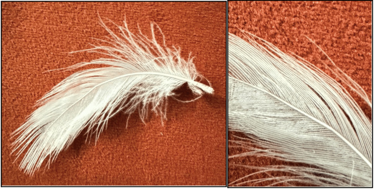 Figure 1
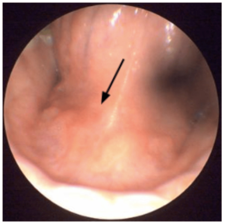 Figure 2
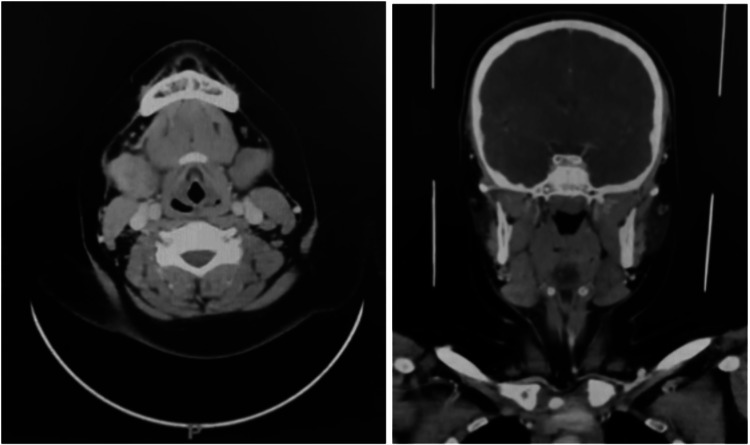 Figure 3
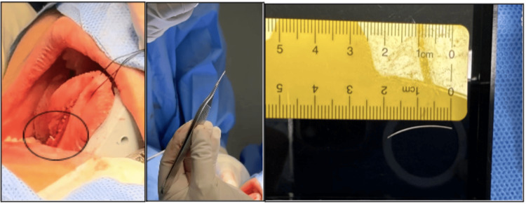 Figure 4
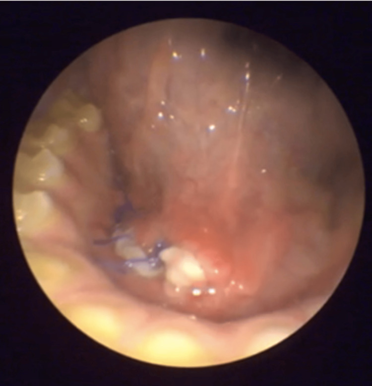 Figure 5
Peer Reviews
No public reviews on file for this paper yet. If you reviewed it on a platform where reviews are public (OpenReview, ICLR, NeurIPS, ICML), you can paste yours below so the community can read it here.
Videos
No videos yet. Explain this paper in a talk, walkthrough, or lecture? Add one.
Taxonomy
TopicsAmerican and British Literature Analysis