Correction: Small molecule treatment alleviates photoreceptor cilia defects in LCA5-deficient human retinal organoids
Dimitra Athanasiou, Tess A. V. Afanasyeva, Niuzheng Chai, Kalliopi Ziaka, Katarina Jovanovic, Rosellina Guarascio, Karsten Boldt, Julio C. Corral-Serrano, Naheed Kanuga, Ronald Roepman, Rob W. J. Collin, Michael E. Cheetham

Abstract
Genes, proteins, chemicals, diseases, species, mutations and cell lines named across the full text — each resolved to its canonical identifier and authoritative record.
Click any figure to enlarge with its caption.
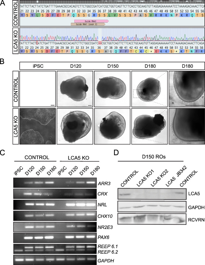 Figure 1
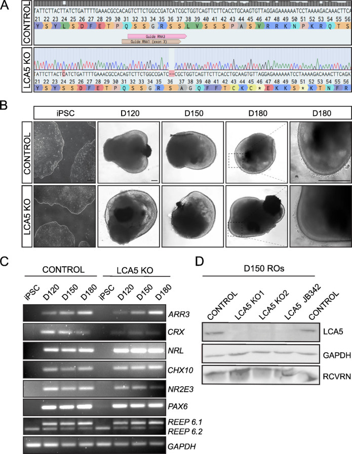 Figure 2
Peer Reviews
No public reviews on file for this paper yet. If you reviewed it on a platform where reviews are public (OpenReview, ICLR, NeurIPS, ICML), you can paste yours below so the community can read it here.
Videos
No videos yet. Explain this paper in a talk, walkthrough, or lecture? Add one.
Taxonomy
TopicsRetinal Development and Disorders · Genetic and Kidney Cyst Diseases
Correction: Acta Neuropathologica Communications (2025) 13:26
10.1186/s40478-025-01943-y
In Fig. 1 of this article [1], an image of iPSC in panel B is missing and have now been corrected in the original publication.
For completeness and transparency, both correct and incorrect versions are displayed below.
Incorrect Fig. 1.
Fig. 1. Generation of LCA5 KO and isogenic control iPSCs and differentiation to retinal organoids. A) Sanger sequence trace of LCA5 KO iPSC (LCA5 KO1) showing a 2-bp deletion in exon 3 of LCA5 gene generated by CRISPR/Cas9 and NHEJ gene editing. B) Bright-field images of iPSC-derived LCA5 KO and isogenic control retinal organoids at D120, D150 and D180 of retinal development. Inset boxes showing the development of photoreceptor brush borders which start to emerge at D180. Scale bars 250 μm. C) RT-PCR of isogenic control and LCA5 KO iPSC and retinal organoids (n = 2 per condition from one differentiation) at D120, D150 and D180 for retinal differentiation markers ARR3, CRX, NRL, CHX10, NR2E3, PAX6, REEP6.1 (upper band), REEP6.2 (lower band). GAPDH was used as a reference transcript. D) Western blot of control, LCA5 KO (KO1 and KO2) and LCA5 JB342 patient retinal organoids at D150 showing successful knockdown of LCA5 protein. Recoverin (RCVRN) was used as a photoreceptor-specific marker and GAPDH as a loading control. Results are from pooling together n = 3 retinal organoids per condition from two differentiations per line
Correct Fig. 1.
Fig. 2. Generation of LCA5 KO and isogenic control iPSCs and differentiation to retinal organoids. A) Sanger sequence trace of LCA5 KO iPSC (LCA5 KO1) showing a 2-bp deletion in exon 3 of LCA5 gene generated by CRISPR/Cas9 and NHEJ gene editing. B) Bright-field images of iPSC-derived LCA5 KO and isogenic control retinal organoids at D120, D150 and D180 of retinal development. Inset boxes showing the development of photoreceptor brush borders which start to emerge at D180. Scale bars 250 μm. C) RT-PCR of isogenic control and LCA5 KO iPSC and retinal organoids (n = 2 per condition from one differentiation) at D120, D150 and D180 for retinal differentiation markers ARR3, CRX, NRL, CHX10, NR2E3, PAX6, REEP6.1 (upper band), REEP6.2 (lower band). GAPDH was used as a reference transcript. D) Western blot of control, LCA5 KO (KO1 and KO2) and LCA5 JB342 patient retinal organoids at D150 showing successful knockdown of LCA5 protein. Recoverin (RCVRN) was used as a photoreceptor-specific marker and GAPDH as a loading control. Results are from pooling together n = 3 retinal organoids per condition from two differentiations per line
The original article has been corrected.