Fresh Upper Gastrointestinal Bleeding (Hematemesis), a Rare Manifestation of Retrograde Intussusception Following Classic Gastric Bypass Surgery
Nader Moeinvaziri, Naser Afshin, Nazanin Setayeshpour, Mohammad Ebrahimi

TL;DR
Upper gastrointestinal bleeding is a rare and potentially misleading complication of retrograde intussusception after gastric bypass surgery.
Contribution
Highlights hematemesis as a rare and under-recognized presentation of retrograde intussusception.
Findings
Retrograde intussusception can present with upper gastrointestinal bleeding, leading to misdiagnosis.
Hematemesis is an uncommon but important clinical manifestation following gastric bypass surgery.
Abstract
Retrograde intussusception may occur following Roux‐en‐Y gastric bypass surgery with a variety of presentations including colicky abdominal pain, constipation, and vomiting. A rare manifestation of this complication is upper gastrointestinal bleeding (hematemesis) which can cause misdiagnosis.
Genes, proteins, chemicals, diseases, species, mutations and cell lines named across the full text — each resolved to its canonical identifier and authoritative record.
Click any figure to enlarge with its caption.
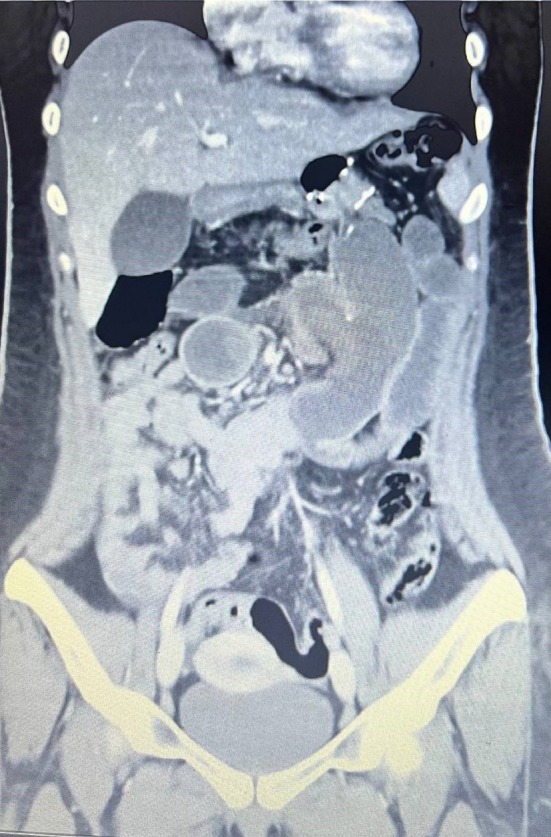 FIGURE 1
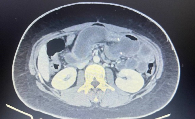 FIGURE 2
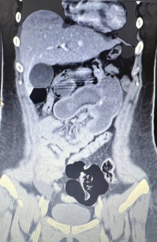 FIGURE 3
Peer Reviews
No public reviews on file for this paper yet. If you reviewed it on a platform where reviews are public (OpenReview, ICLR, NeurIPS, ICML), you can paste yours below so the community can read it here.
Videos
No videos yet. Explain this paper in a talk, walkthrough, or lecture? Add one.
Taxonomy
TopicsGastrointestinal disorders and treatments · Gastrointestinal Bleeding Diagnosis and Treatment · Gastrointestinal Tumor Research and Treatment
Introduction
1
Bariatric surgery (BS) is considered the most efficient long‐term therapy for morbid obesity [1]. Laparoscopic Roux‐en‐Y gastric bypass (RYGB) is generally considered a safe and effective procedure to prevent long‐term complications of morbid obesity [2]. Some complications such as bleeding, anastomosis stricture, marginal ulcers, leakage from the anastomosis site, and intussusception have been reported after gastric bypass [2, 3].
Intussusception is rare in adults, accounting for just 5% of all cases, and 90% of them, are due to an underlying pathology (like tumors or inflammatory processes, and polyps) acting as a leading point, causing invagination of the proximal part of the bowel into the distal alongside peristalsis (anterograde) [4]. Retrograde intussusception mostly follows Roux‐en‐Y gastric bypass [5]. In the RYGB, the distal bowel (common channel) is drawn into the lumen of the proximal bowel (alimentary limb or jejunojejunostomy anastomosis). These intussusceptions usually occur without a leading point and may represent a motility disorder following the Roux‐en‐Y reconstruction, which may require surgical intervention [6].
Diagnosis of intussusception is difficult due to its presentation with a wide range of symptoms and various severities, including acute or chronic abdominal pain and obstructive symptoms, and laboratory test results are not specific [7, 8]. In this study, we represent a 19‐year‐old woman with an unusual presentation of intussusception presenting as upper gastrointestinal bleeding (Hematemesis), which can cause misdiagnosis.
Case Presentation
1.1
A 19‐year‐old woman with a body mass index (BMI) of 47 (137 kg weight and 172 cm height) underwent RYGB surgery. The procedure was done by a standard method with 5 ports. A gastric pouch was created by three 60 mm Endo‐GI purple staplers, a gastrojejunal anastomosis was about 2 cm with an alimentary limb of 130 cm, and a biliopancreatic limb of 70 cm. Peterson and jejunojejunal defects were closed completely.
Diet was started for her the next day, and she was discharged the second day after surgery in a stable condition with instructions to have regular postoperation follow‐ups. She weighed 109 kg and 93 kg at 3 and 6 months postoperation follow‐ups, respectively. Iron deficiency anemia was detected in routine lab tests, and iron supplements were prescribed for her. Following her 6‐month follow‐up, she presented with low‐intensity colicky abdominal pain episodes after food ingestion that resolved spontaneously after a few minutes. However, she neglected the painful episodes as they were self‐limited.
Nine months after the surgery, she was admitted to the hospital due to 2 episodes of massive bloody vomiting and abdominal pain (worse than her previous pains). Her vital signs were stable upon admission, and she had a low‐intensity colicky pain in the left upper quadrant without tenderness. She was scheduled for an upper gastrointestinal endoscopy, which showed a marginal ulcer at the site of the gastrojejunostomy anastomosis without active bleeding. The next day, her abdominal pain worsened along with upper abdominal tenderness during her physical examination. Her laboratory data showed elevated white blood cells from 8500 to 12,000 / μL and a mild decrease in hemoglobin concentration from 11 to 10.5 g/dL. An abdominal and pelvic computed tomography (CT scan) with contrast was done, which showed distention in small bowel lumen with a target sign (Figures 1, 2, 3).
Abdominal CT demonstrating the target sign of a small‐bowel intussusception.
Axial abdominal CT demonstrating the classic target/bull's‐eye sign.
Coronal view of small bowel intussusception.
With suspicion of intestinal obstruction and intussusception, the patient underwent laparotomy. Her surgical findings were retrograde intussusception of the common channel at the jejunojejunal anastomosis site with severe edema of the small intestine, which made bowel reduction impossible due to patchy areas of necrosis in the intestine. Consequently, the beginning of the common channel and the jejunojejunal anastomosis sites were resected, and Roux‐en‐Y configuration was re‐established. First, the end of the common channel anastomosed to the Roux limb, then the biliopancreatic limb connected to the common channel 30 cm distally, both in side‐toto‐side manner.
Outcome and Follow‐up
2
Her vital signs were stable 3 days after the surgery, and she tolerated a liquid diet after a normal dye study. On the fifth day after the operation, the drain was removed, and the patient was discharged in excellent condition. Two weeks after surgery, the patient visited without any definite complaints. she also visited after 3 months for her routine follow‐up, which had 6 kg weight loss and acceptable laboratory parameters.
Conclusion
3
In conclusion, a rare complication following Roux‐en‐Y gastric bypass surgery is retrograde intussusception with a variety of presentations including colicky abdominal pain, constipation, nausea, and vomiting, and even upper gastrointestinal bleeding (hematemesis) just like our case that can cause misdiagnosis during management. We believe that this paper is the first jejunojejunal intussusception presenting itself with hematemesis that is reported in the literature following RYGB surgery. Clinicians should suspect the presence of intussusception after RYGB even with fresh upper gastrointestinal bleeding presentation along with abdominal pain.
Discussion
4
Intussusception is rare in adults, and retrograde intussusception is even less common, with a variety of nonspecific symptoms including bowel obstruction, constipation, abdominal distention, and more commonly, abdominal pain, causing the diagnosis to be a challenging endeavor [7, 9, 10]. The usual symptoms that patients present with are epigastric pain with discrete intensity and duration, nausea, and vomiting [9]. In addition, rare cases present with a triad of abdominal pain, hematochezia, and an abdominal mass [4]. Post‐RYGB transient colicky abdominal pain should be considered important, as it may be an early presentation of intussusception or internal hernias. Our patient complained of recurrent limited abdominal pain, which, if presented sooner, could have been prevented by earlier intervention from progressing to complications and necrosis. Leading points such as tumors or inflammatory processes and polyps are known etiologies of intussusception in adults. however, some other causes such as significant weight loss, disruption of the duodenal pacemaker post transection of the jejunum, dysmotility after RYGB, and existence of the stapler line as a leading point may predispose the patients post RYGB to intussusception [11] Branch et al. reported a Roux‐en‐Y gastric bypass patient with jejunojejunostomy intussusception who also had hematemesis; though in that case, a concurrent bleeding marginal ulcer was found to be the source [12] There has been a reported case of hematemesis post invagination of the efferent limb into the gastrojejunostomy anastomosis in a patient with a previous history of billroth II surgery due to a complicated peptic ulcer [13] To the best of our knowledge, this is the first reported jejunojejunal intussusception presenting itself with upper GI bleeding. The patient had no complaints of hematemesis or melena during the entire 9 months after the operation until the time of hospitalization and only complained of colicky abdominal pain. During the endoscopy procedure, no evidence of active bleeding was detected at the anastomotic site. Also, during surgery and bowel resection, the secretions inside the intestine were bloody. We hypothesize that, due to the obstruction distal to the jejunojejunal anastomosis, the bloody secretions moved towards the alimentary limb and manifested as hematemesis. Given the above evidence, we assume that the bleeding is caused by vascular congestion, mucosal ischemia, and hemorrhage within the small intestine following intussusception. Although examination and laboratory data do not specify the diagnosis and ultrasound usually is not helpful in adult cases of intussusception, a CT scan with contrast can be the best modality for such cases, showing “target sign” which is suggestive of intussusception diagnosis [9, 14]. The pathology of the resected bowel also was in favor of transmural necrosis without any definite underlying cause.
The decision of surgical or conservational management of these cases depends on the patient's condition and the complications that follow. Some cases have shown a spontaneous reduction of intussusception; however, in other cases, bowel blockage, bowel ischemia, and bowel distention have been reported, causing necessary and emergency surgical management [9]. The challenging choice for the management of such cases can be eased with contrast abdominal CT scans [14]. Different surgical management choices have been suggested in the literature for intussusception, including intussusception reduction and resection of the damaged bowel with re‐establishing anastomosis [15]. Although some studies have reported enteropexy or mesenteric fixation to prevent the intussusception post RYGB [8], no definite recommended method for prevention has been reported to date. Ankur Makani et al. reported a case of recurrent intussusception post RYGB, which underscores the importance of understanding the causes for better diagnosis and prevention [11].
Author Contributions
Naser Afshin: conceptualization, supervision, writing – original draft. Nazanin Setayeshpour: writing – review and editing. Mohammad Ebrahimi: data curation, resources. Nader Moeinvaziri: project administration, supervision, writing – original draft, writing – review and editing.
Consent
Written informed consent was obtained from the patient and her family.
Conflicts of Interest
The authors declare no conflicts of interest.
The reference list from the paper itself. Each links out to its DOI / PubMed record.
- 1S. Singla , B. A. Guenthart , L. May , J. Gaughan , and J. E. Meilahn , “Intussusception After Laparoscopic Gastric Bypass Surgery: An Underrecognized Complication,” Minimally Invasive Surgery 2012, no. 1 (2012): 464853, 10.1155/2012/464853.22991661 PMC 3444049 · doi ↗ · pubmed ↗
- 2S. K. Dutta , M. Arora , A. Kireet , H. Bashandy , and A. Gandsas , “Upper Gastrointestinal Symptoms and Associated Disorders in Morbidly Obese Patients: A Prospective Study,” Digestive Diseases and Sciences 54 (2009): 1243–1246, 10.1007/s 10620-008-0485-6.18975090 · doi ↗ · pubmed ↗
- 3G. Orthopoulos , H. M. Grant , P. Sharma , E. Thompson , and J. R. Romanelli , “S 054: Incidence and Management of Jejunojejunal Intussusception After Roux‐En‐Y Gastric Bypass: A Large Case Series,” Surgical Endoscopy 34 (2020): 2204–2210, 10.1007/s 00464-019-07009-0.31385075 · doi ↗ · pubmed ↗
- 4D. Sarma , R. Prabhu , and G. Rodrigues , “Adult Intussusception: A Six‐Year Experience at a Single Center,” Annals of Gastroenterology 25, no. 2 (2012): 128–132.24714146 PMC 3959399 · pubmed ↗
- 5L. Daellenbach and M. Suter , “Jejunojejunal Intussusception After Roux‐En‐Y Gastric Bypass: A Review,” Obesity Surgery 21 (2011): 253–263, 10.1007/s 11695-010-0298-5.20949329 · doi ↗ · pubmed ↗
- 6F. C. Brunicardi , D. K. Andersen , T. R. Billiar , et al., Schwartz's Principles of Surgery 2‐Volume Set, 11th ed. (Mc Graw Hill Professional, 2019).
- 7G. Kasotakis and R. Sudan , “Retrograde Intussusception After Roux‐En‐Y Gastric Bypass for Morbid Obesity,” Obesity Surgery 19 (2009): 381–384, 10.1007/s 11695-008-9775-5.19067088 · doi ↗ · pubmed ↗
- 8C. L. Kalmar and T. A. Lucktong , “Laparoscopic Enteropexy for Intussusception at Roux‐En‐Y Anastomosis,” Journal of Surgical Case Reports 5 (2019): rjz 152, 10.1093/jscr/rjz 152.PMC 651643131110656 · doi ↗ · pubmed ↗