A Case of Successful Extraction of a Micra Leadless Pacemaker Implanted Two Years Prior Using the Axis-Guided Dual Snare Technique
Misun Pak, Ken Kakita, Takashi Yamasaki, Tetsuhisa Hattori

TL;DR
A 78-year-old man's Micra pacemaker was successfully extracted using a new dual snare technique after causing heart issues.
Contribution
A novel axis-guided dual snare technique was successfully used for extracting a Micra leadless pacemaker.
Findings
The dual snare technique successfully stabilized and extracted the Micra device.
The patient's heart condition improved after the extraction and subsequent CRT implantation.
Abstract
A 78-year-old man underwent Micra AV implantation due to complete atrioventricular block. He developed diffuse left ventricular systolic dysfunction and dyssynchrony, 2 years later, suggesting pacing-induced cardiomyopathy. Given the critical need for an upgrade to cardiac resynchronization therapy (CRT), an extraction of the Micra AV was scheduled. Initially, the Micra extraction using a solitary snare catheter was attempted, but capturing the retrieval feature on the proximal side of the device was unsuccessful due to excessive movement caused by the heartbeats. Consequently, an axis-guided dual snare technique employing two snare catheters was implemented. A triple-loop snare catheter was inserted into a steerable sheath, which was threaded through the loop of a single-loop snare catheter placed outside the sheath. The triple-loop snare successfully grasped the Micra body and…
Genes, proteins, chemicals, diseases, species, mutations and cell lines named across the full text — each resolved to its canonical identifier and authoritative record.
Click any figure to enlarge with its caption.
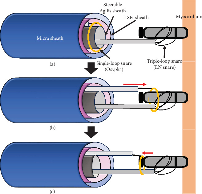 Figure 1
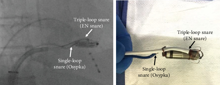 Figure 2
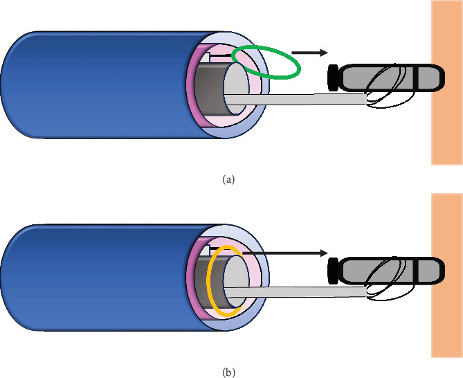 Figure 3
Peer Reviews
No public reviews on file for this paper yet. If you reviewed it on a platform where reviews are public (OpenReview, ICLR, NeurIPS, ICML), you can paste yours below so the community can read it here.
Videos
No videos yet. Explain this paper in a talk, walkthrough, or lecture? Add one.
Taxonomy
TopicsCardiac pacing and defibrillation studies · Advanced MRI Techniques and Applications · Cardiac Arrhythmias and Treatments
1. Introduction
The Micra transcatheter pacing system (Micra TPS, Medtronic, Minneapolis, Minnesota) is a leadless pacemaker that offers several advantages over traditional transvenous pacemakers, including resistance to infections and avoidance of venous occlusions [1, 2]. A Micra extraction is sometimes necessary due to elevated pacing thresholds or the need to upgrade to a cardiac resynchronization therapy (CRT) device. Generally, the outcomes of Micra extractions in the chronic phase are relatively acceptable, and some successful cases of late Micra retrievals have been reported [3–5]. For a safe device removal, it is crucial to grasp the neck of the retrieval feature on the proximal side of the Micra [6, 7]. Although snaring the neck of the retrieval feature is necessary for extraction, its small structure requires precise and delicate snare manipulation. This is particularly challenging when the Micra moves significantly due to heartbeats or when the device is encapsulated by a fibrous tissue [8–11]. This case report details the successful extraction of a Micra implanted 2 years prior by the axis-guided dual snare technique, which smoothly captured the retrieval feature using two snare catheters.
2. Case Presentation
A 78-year-old man underwent a Micra AV implantation due to complete atrioventricular block. A Micra AV was implanted on the right ventricular (RV) mid-septum, and all electrical parameters were acceptable. However, he developed heart failure with a reduced left ventricular ejection fraction dropping from 60% before the implantation to 35% 2 years later. Transthoracic echocardiography revealed left ventricular dyssynchrony. The possibility of cardiomyopathies, including ischemic heart disease, was ruled out, and he was finally diagnosed with pacing-induced cardiomyopathy (PICM). Consequently, an upgrade to a CRT device was deemed essential, and the Micra extraction was scheduled.
The Micra extraction was performed under general anesthesia in preparation for any emergent surgeries. Initially, a Micra TPS introducer sheath (outer diameter: 27-French, Medtronic, Minneapolis, Minnesota) was inserted via the right femoral vein. To prevent back bleeding, a short sheath (outer diameter: 18-French) was introduced into the Micra sheath, followed by a steerable sheath (Agilis NXT, outer diameter: 8.5-French, Abbott, Santa Clara, California) (sheath-in-sheath technique) [12]. We initially attempted its removal using a solitary snare catheter. A triple-loop EN Snare Endovascular Snare System (Merit Medical System, South Jordan, Utah) was advanced through the steerable sheath to capture the retrieval feature on the proximal side of the Micra. However, grasping the small retrieval feature proved challenging due to the device's significant pivotal motion caused by the heartbeats, despite the triple-loop snare successfully grasping the Micra body.
After spending over 30 min trying to capture the retrieval feature by a solitary snare catheter, a Micra extraction using two snare catheters was subsequently attempted. Initially, a triple-loop EN snare catheter was inserted into a steerable Agilis sheath, which was threaded through a loop of a single-loop snare catheter (Osypka Medical GmBH, Berlin, Germany) placed outside the sheath. This system was inserted together into the 18-Fr short sheath (Figure 1a). The triple-loop EN snare successfully grasped the Micra body and stabilized its excessive movement. Following this, the single-loop Osypka snare could be advanced along the shaft of the triple-loop EN Snare towards the Micra body (Figure 1b). The single-loop Osypka snare was gradually pulled back from the body to the proximal side of the Micra while its loop was made smaller, ultimately capturing the neck of the retrieval feature and the shaft of the EN Snare simultaneously (Figure 1c). By holding both the Micra body and retrieval feature with the two snare catheters, the alignment of the Micra and the steerable Agilis sheath could be maintained coaxially, which facilitated a smooth retrieval into the Micra sheath (axis-guided dual snare method) (Figure 2). It took only about 5 min to insert two snares into the sheath, extract the Micra, and pull back it into the sheath. After the Micra extraction, a CRT device was implanted without any complications.
3. Discussion
In this case, a Micra AV implanted 2 years prior was successfully removed using an axis-guided dual snare technique. This technique enabled operators to easily capture the Micra's retrieval feature by advancing one snare along the axis of another snare that was grasping the Micra's body.
For successful Micra extractions in the chronic phase, the double-snare technique has been previously reported. This method involves firmly grasping the Micra's body with the initial snare and catching the retrieval feature with the second snare, which assists in device extraction [6]. However, this technique still requires the additional effort of capturing the small retrieval feature with the second snare. It may be hard to capture the retrieval feature from scratch, even if excessive movement of the Micra can be controlled with the initial snare (Figure 3a). On the other hand, the axis-guided dual snare technique is considered to be more effective for Micra extractions compared to the conventional approach. The key point of this technique is that the initial snare catheter is threaded through the loop of the second snare in advance. Consequently, the retrieval feature can be easily captured by simply advancing the second snare along the shaft of the initial snare holding the Micra body (“axis-guided”), eliminating the need for the burdensome manipulation of the second snare to catch the retrieval feature (Figure 3b). In this case, the solitary snare method failed to capture the retrieval feature despite significant effort. On the other hand, the axis-guided dual snare technique proved to be faster and more effective, simplifying the procedure. Since device extraction is often followed by the implantation of a new pacemaker or CRT, reducing the procedure time benefits both patients and operators. Therefore, this method is a practical and reliable first-choice approach for Micra extraction.
In our method, the Micra was removed by simultaneously capturing both the retrieval feature and the shaft of the En snare holding the Micra body with the Osypka snare catheter. Ideally, grasping only the retrieval feature is preferable in Micra retrieval. However, it was assumed that the Micra could be reliably extracted due to the strong gripping force of the Osypka snare catheter, even when both the retrieval feature and the shaft of the En snare were grasped simultaneously [13].
This case report highlights the utility of the axis-guided dual snare technique. However, its feasibility and reproducibility are uncertain, necessitating further studies with larger datasets. Additional data would help confirm the consistency and broader applicability of this technique. Furthermore, even with advancements in Micra extraction techniques, the extraction procedures are recommended to be performed in facilities equipped with cardiac surgical teams to address potential complications such as cardiac perforation or accidental dislodgments of the Micra during the extraction procedure [14, 15].
4. Conclusion
A Micra implanted 2 years prior was completely extracted using the axis-guided dual snare technique. This method can be considered as a potential first-choice for Micra extraction.
The reference list from the paper itself. Each links out to its DOI / PubMed record.
- 1Reynolds D. Duray G. Z. Omar R. A Leadless Intracardiac Transcatheter Pacing System The New England Journal of Medicine 2016374653354110.1056/NEJ Moa 15116432-s 2.0-8495927653726551877 · doi ↗ · pubmed ↗
- 2Okabe T. Afzal M. R. Houmsse M. Tine-Based Leadless Pacemaker Strategies for Safe Implantation in Unconventional Clinical Scenarios JACC: Clinical Electrophysiology 20206101318133110.1016/j.jacep.2020.08.02133092762 · doi ↗ · pubmed ↗
- 3Kancharla K. Deshmukh A. J. Friedman P. A. Leadless Pacemakers - Implant, Explant and Long-Term Safety and Efficacy Data Journal of Atrial Fibrillation 20171021610.4022/jafib.15812-s 2.0-8503465725129250229 PMC 5673288 · doi ↗ · pubmed ↗
- 4Minami K. Petru J. Chovanec M. Late Retrieval of the Leadless Micra Transcatheter Pacemaker System Journal of Clinical Cardiology and Cardiovascular Interventions 20214611310.31579/2641-0419/138 · doi ↗
- 5Grubman E. Ritter P. Ellis C. R. To Retrieve, or Not to Retrieve: System Revisions With the Micra Transcatheter Pacemaker Heart Rhythm 201714121801180610.1016/j.hrthm.2017.07.0152-s 2.0-8503714534428713024 · doi ↗ · pubmed ↗
- 6Goyal R. Markowitz S. M. Ip J. E. Double-Snare Technique for Capturing a Wandering Leadless Pacemaker JACC: Clinical Electrophysiology 20195787287310.1016/j.jacep.2019.02.0102-s 2.0-8506821660431320021 · doi ↗ · pubmed ↗
- 7Li J. Hou W.-B. Cao M. K. Safety and Efficacy of Leadless Pacemaker Retrieval Journal of Cardiovascular Electrophysiology 20193091671167810.1111/jce.140762-s 2.0-8507178229631310416 · doi ↗ · pubmed ↗
- 8Kypta A. Blessberger H. Lichtenauer M. Steinwender C. Complete Encapsulation of a Leadless Cardiac Pacemaker Clinical Research in Cardiology 20161051 p. 9410.1007/s 00392-015-0929-x 2-s 2.0-8495519387026493306 · doi ↗ · pubmed ↗