HDAC11 promotes renal fibrosis by induing partial epithelial-mesenchymal transition and G2/M phase arrest in renal epithelial cells
Yingjie Guan, Fengchen Shen, Liyuan Yao, Ting C. Zhao, Shougang Zhuang

TL;DR
This study shows that HDAC11 contributes to kidney scarring by causing cell changes and cell cycle arrest, suggesting it could be a target for treatment.
Contribution
The study identifies HDAC11 as a novel driver of renal fibrosis through partial EMT and G2/M arrest mechanisms.
Findings
HDAC11 inhibition reduced collagen accumulation and fibrosis markers in mouse models.
HDAC11 suppression decreased pro-inflammatory cytokines and macrophage infiltration in obstructed kidneys.
Pharmacological and genetic HDAC11 inhibition suppressed profibrotic signaling pathways like Smad3, STAT3, and NF-κB.
Abstract
Histone deacetylase 11 (HDAC11) is the sole member of class IV HDACs, implicated in tumor growth, immune regulation, and oxidative stress injury. Its specific role in renal fibrosis and underlying mechanisms remains unclear. The global knockout of HDAC11 mice and FT895, a selective inhibitor of HDAC11, were utilized to assess the role of HDAC11 in renal fibrosis following unilateral ureteral obstruction (UUO) injury in mice. Immunostaining was employed to analyze renal expression of HDAC11 and infiltration of macrophages. Immunoblot analysis was used to analyze the expression and/or phosphorylation of proteins associated with partial epithelial-mesenchymal transition (pEMT) in the kidney and cultured renal proximal tubular cells (RTPCs). RT-PCR was used to analyze the expression of various proinflammatory cytokines. HDAC11 was predominantly expressed in renal epithelial cells, with…
Genes, proteins, chemicals, diseases, species, mutations and cell lines named across the full text — each resolved to its canonical identifier and authoritative record.
Click any figure to enlarge with its caption.
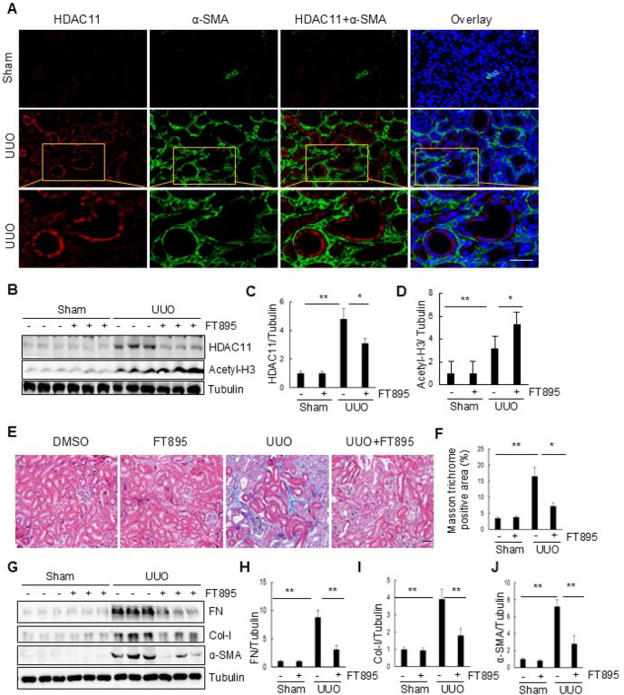 Figure 1
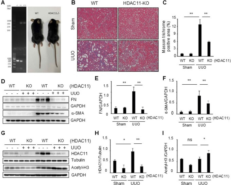 Figure 2
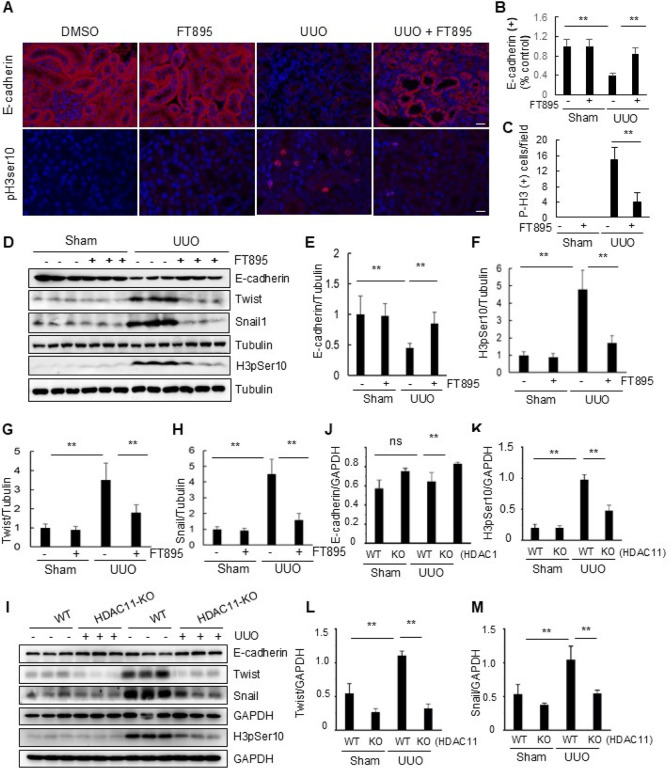 Figure 3
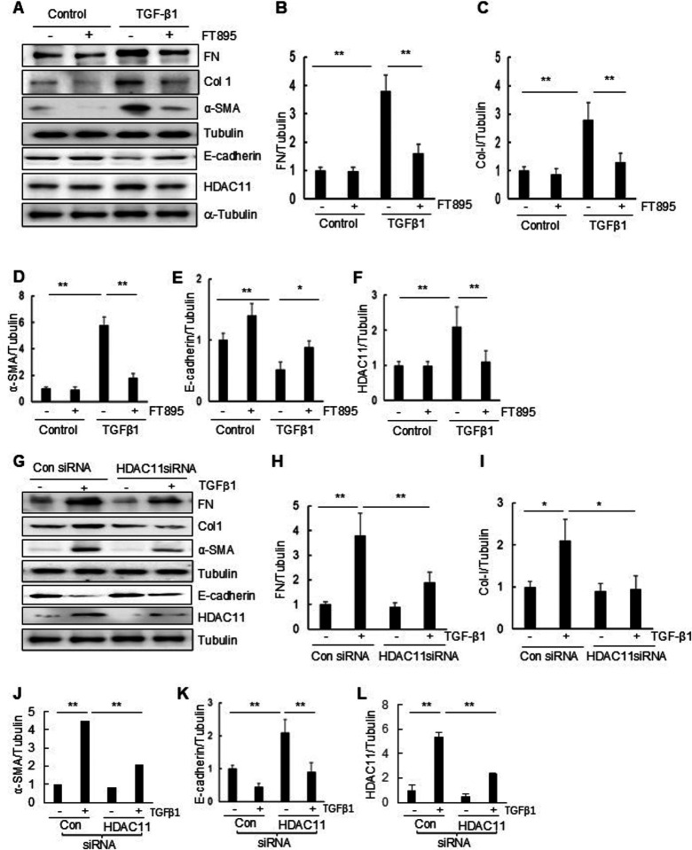 Figure 4
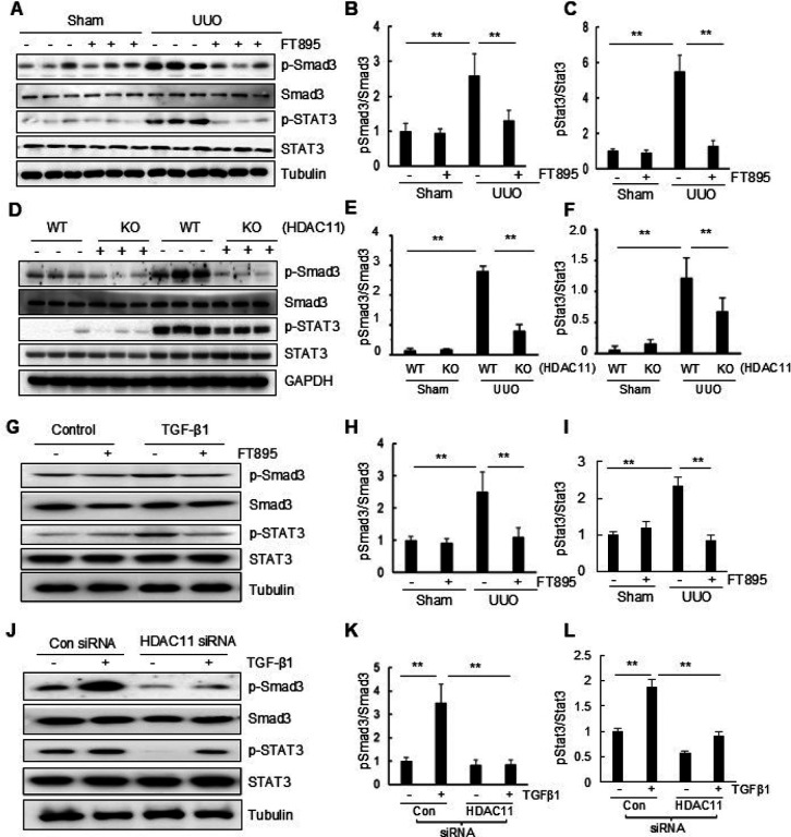 Figure 5
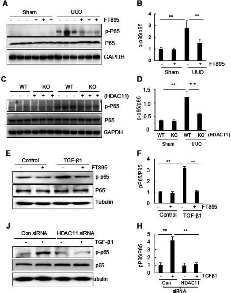 Figure 6
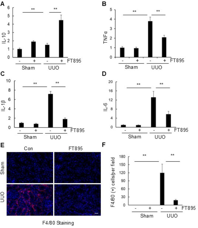 Figure 7
Peer Reviews
No public reviews on file for this paper yet. If you reviewed it on a platform where reviews are public (OpenReview, ICLR, NeurIPS, ICML), you can paste yours below so the community can read it here.
Videos
No videos yet. Explain this paper in a talk, walkthrough, or lecture? Add one.
Taxonomy
TopicsHistone Deacetylase Inhibitors Research · Epigenetics and DNA Methylation · Adenosine and Purinergic Signaling
Background
Chronic kidney disease (CKD) is a global public health problem. It affects more than 20 million people in the United States alone.(Lerner et al., 2017) Regardless of the etiologies, CKD would progress to end stage of renal disease.(Bello et al., 2024; Lerner et al., 2017) In the past several decades, studies have revealed that the common pathway underlying CKD is kidney fibrosis, which is characterized by renal interstitial fibroblast activation, extracellular matrix (ECM) accumulation, inflammatory responses and destruction of normal structure.(Bello et al., 2024; Reiss et al., 2024) Currently, there is still no specific treatment for targeting fibrosis. This enormous unmet medical need calls for better understanding of the mechanism underlying fibrotic CKD.
Increasing evidence has revealed that partial epithelial-mesenchymal transition (pEMT) is a critical pathological process that initiates activation of renal interstitial fibroblasts and production of ECM components in the kidney following various injuries.(Lovisa et al., 2016; Sheng and Zhuang, 2020) pEMT is defined as the state in which epithelial cells express markers of both epithelial and mesenchymal cells, but remain associated with the basement membrane.(Lovisa et al., 2016; Sheng and Zhuang, 2020) The renal epithelial cells with pEMT are arrested at the G_2_/M phase of cell cycle, and able to generate a large amount of profibrotic growth factors/proinflammatory factors. Subsequently, these factors are released into the renal interstitium where induces the transformation of fibroblasts to active fibroblasts (myofibroblasts).(Lovisa et al., 2016; Sheng and Zhuang, 2020) Several signaling pathways such as transforming growth factor-b/Smad3 (TGF-b/Smad3), signal transducer and activator of transcription 3 (STAT3) and some transcription factors such as Snail and Twist, have been identified to participate in the process of pEMT in the injured kidney.(Lovisa et al., 2016; Pang et al., 2010; Sheng and Zhuang, 2020) Recently, a number of post-translational modifications, including protein acetylation, have also been recognized to contribute to this process.
Protein acetylation is one of the post-translational modifications that occurs in both histone and non-histone proteins.(Shen and Zhuang, 2022) Histone acetylation often occurs at positively charged lysine residues which weakens the DNA-histone interactions, thus opening the chromatin and facilitating transcription(Shen and Zhuang, 2022). For example, acetylation of lysine 9 on histone 3 (H3K9ac) correlates with transcription activation.(Nozaki and Kanai, 2021) Acetylation also involves in regulation of many non-histone proteins, including transcription factors, transcriptional coactivators and nuclear receptors(Shvedunova and Akhtar, 2022). Acetylation is positively regulated by histone acetyltransferases and negatively by histone deacetylates.(Shen and Zhuang, 2022; Wang et al., 2022) As such, either activation of histone acetyltransferases or inactivation of histone deacetylates is able to promote protein acetylation.(Shen and Zhuang, 2022) To date, numerous histone acetyltransferases and histone deacetylates have been identified. Depending on their sequence homology, HDACs are divided into four categories with 11 isoforms: class I HDACs (HDAC1–3,8), class II HDACs (HDAC 4–7, 9; 10), class III [sirtuin (SIRT)1–SIRT7], and class IV (HDAC11). The various HDACs exhibit distinct functions, unique cellular and organ distribution, as well as discrete physiological and pathological effects(Shen and Zhuang, 2022; Tang and Zhuang, 2019).
Previous studies have shown that HDACs play a key role in the development of various kidney diseases. (Shen and Zhuang, 2022) Certain HDAC isoforms, such as HDAC3, 4, 6, 8 and 9, are upregulated in injured kidneys and contribute to renal fibrosis.(Chen et al., 2020; Pang et al., 2009; Shen and Zhuang, 2022; Zhang et al., 2020) These findings were obtained using either class- or isoform-specific inhibitors or conditional deletion techniques.(Shen and Zhuang, 2022) A recent study showed that inhibition of HDAC11 by quisinostat attenuated renal fibrosis induced by unilateral ureteral obstruction (UUO).(Mao et al., 2020) However, quisinostat is not a specific HDAC11 inhibitor and can also target several other HDACs with equivalent potency - all of which have been shown to play regulatory roles in renal fibrosis, (Arts et al., 2009) thus, it remains uncertain about the role and mechanisms of HDAC11 in renal fibrosis.
In this study, we employed globally deleted HDAC11 mice and FT895, a highly selective inhibitor of HDAC11,(Martin et al., 2018) to investigate the role and mechanism of HDAC11 in renal fibrosis. Our results demonstrate that either global deletion of HDAC11 or administration of FT895, attenuated renal fibrosis, pEMT and G2/M phase arrest in renal epithelial cells in a murine model of UUO. Both FT895 treatment and siRNA-mediated silencing of HDAC11 also inhibited TGF-b1 induced pEMT in cultured murine renal proximal tubular epithelial cells (RPTCs). Based on the data, we concluded that HDAC11 is critically involved in renal fibrogenesis through a mechanism involving the suppression of EMT and G2/M phase arrest in renal epithelial cells.
METHODS
Chemicals and antibodies
Antibodies for collagen I (A2), snail, twist, and glyceraldehyde 3-phosphate dehydrogenase (GAPDH) were sourced from Santa Cruz Biotechnology (Dallas, TX, USA). Antibodies to phospho-histone H3 at Ser10 (pH3Ser10) (for immunoblot analyses), acetylated Histone 3 antibodies, α-smooth muscle actin (α-SMA), and α-tubulin were purchased from Millipore Sigma (Burlington, MA, USA). Additionally, antibodies to fibronectin, HDAC11, phospho-Smad3, and Smad3 antibodies along with siRNA specific for HDAC11 were obtained from Invitrogen (MA, USA). FT895 was acquired from Cayman (MA, USA). Anti–F4/80 was purchased from Abcam (Waltham, MA, USA). All other items, including antibodies to phospho-STAT3, STAT3, phospho-NF-KB(p65) NF-κB(p65), snail1, pH3Ser10 (for immunofluorescence staining) or E-cadherin were obtained from Cell Signaling Technology (Danvers MA, USA);.
Animals and experimental design
Global HDAC11 knockout mice (HDAC11^−/−^) were obtained from Shanghai Model Organism. These mice were found to be viable and developed normally, as reported in previous studies.(Nunez-Alvarez et al., 2021) The UUO model was established in male C57B6 black mice weighing 20–25 g (The Jackson Laboratory, Bar Harbor, ME, USA) in accordance with our previous studies(Pang et al., 2009). In brief, a flank incision was made to expose the abdominal cavity, and the left ureter was isolated and ligated. The contralateral kidney served as a control. To investigate the role of HDAC11 in renal fibrosis, FT895 (5 mg/kg) in 50 μl DMSO was immediately administered by i.p. injection after ureteral ligation and then given daily for 6 days. The dosage of HDAC11 was chosen based on a previous report by Rau et al(Martin et al., 2018). For the UUO-alone group, mice were injected with an equivalent amount of DMSO. At 7 days post-ligation, mice were euthanized and both obstructed and contralateral non-obstructed kidneys were harvested for various analysis. All animal study procedures were approved by the Lifespan Institutional Animal Care and Use Committee and conducted in compliance with the NIH Guide for the Care and Use of Laboratory Animals. The animals had ad libitum access to standard rodent chow and water.
Histology and Immunohistochemistry Analysis
Mouse kidney samples were fixed in 10% formalin (Sigma-Aldrich) overnight and embedded in paraffin. 5-μm thick sections were used for immunohistochemistry. Masson trichrome staining was carried out according to the procedure described in previous studies(Pang et al., 2010). To quantitatively assess renal fibrosis, the collagen tissue area was measured using Image Pro-Plus Software (Media Cybernetics, Rockville, MD, USA) by drawing a line around the perimeter of the positive staining area, and calculating and graphing the mean ratio to each microscopic field. For immunofluorescence staining, sections were deparaffinized, rehydrated and antigen retrieved at 98°C for 10 minutes in 10 mM citrate buffer pH 6. The tissue sections were incubated with 5% normal goat serum (NGS), 5% BSA in TBST for 30 minutes prior to overnight incubation with the primary antibody. The following primary antibodies were used: anti–phospho–histone H3 (Abcam, 1:3,000), anti–mouse HDAC11 (Santa Cruz, 1:100), anti–mouse α-SMA (Sigma, 1:1000), anti–rabbit E cadherin (cell signal, 1:100), and anti–rat F4/80 (Abcam, 1:200). 4’,6-diamidino-2-phenylindole (DAPI) was used to stain nuclei. Eight to ten randomly selected fields at a magnification of 400× on each section were then digitally photographed and scored in a blinded fashion via computerized morphometric analysis.
Western blot analysis
Cell and mouse kidneys were homogenized in RIPA lysis buffer (150 mM NaCl, 50 mM Tris–HCl pH 8, 1% NP-40, 0.5% sodium deoxycholate, 0.1% SDS) supplemented with a protease inhibitor cocktail (Complete^™^, Roche). The total protein amount was determined using the BCA assay (Thermo Scientific), and equal amounts of lysates for each sample were separated by SDS-PAGE and transferred to nitrocellulose membranes. Following a 1-hour incubation with 5% nonfat milk at room temperature, the membranes were then exposed to a primary antibody overnight at 4°C followed by an appropriate horseradish peroxidase-conjugated secondary antibody for 1 hour. Bound antibodies were visualized using chemiluminescence detection. The semi-quantitative analysis of different proteins was conducted using ImageJ software (NIH). Densitometry analyses were quantified based on the intensity (density) of the band, which was calculated by the area and pixel value of the band. The quantification data are presented as a ratio between the target protein and loading control (housekeeping protein).
Quantitative real–time PCR analysis
RNA extraction and quantitative real-time polymerase chain reaction (PCR) were conducted following the procedure outlined in our previous studies(Wang et al., 2018). Kidneys were homogenized, and total RNA was extracted according to the manufacturer’s instructions (Qiagen). cDNA synthesis was carried out using the Bio-Rad cDNA synthesis kit as per the manufacturer’s protocol. The mRNA expression levels of the listed genes were quantified by real-time PCR using the SYBR Green PCR Master mix (Qiagen) and normalized to the expression of GAPDH housekeeping gene. Relative transcription levels were calculated as previously described(Pang et al., 2010). The genes and primer sequences are identical to those previously utilized(Pang et al., 2010).
Cell culture and treatment
Mouse renal proximal tubular epithelial cells (RPTCs) were cultured in DMEM medium supplemented with 10% heat–inactivated fetal bovine serum and penicillin/streptomycin (100 ~ g/ml) at 37°C and 5% CO2. To assess the impact of FT895, various doses of FT895 were directly added to the sub-confluent RPTCs, followed by a 24-hour incubation period. For the induction of EMT, RPTCs were starved for 24 hours in serum-free medium and then exposed to 5 ng/ml TGF-β1 (R&D Systems) for 48 hours in the presence or absence of FT895. The control for TGF-β1 treatment utilized a vehicle consisting of 4 mM HCl in H2O with 1 mg/ml BSA. To transfect siRNA into RPTC cells, we seeded cells to a confluence of 60–70% in antibiotic-free medium and transfected them with siRNA specific to HDAC11 (100 pmol) using Lipofectamine 2000 (Thermo Fisher Scientific, Waltham, MA, USA), following the manufacturer’s instructions. In parallel, scrambled siRNA (100 pmol) was used as a control for off-target changes in RPTC cells. Twenty-four hours after transfection, the medium was changed and cells were incubated in the absence or presence of TGF-b1 for an additional 24 hours before being harvested for analysis.
Statistical analyses
The data are presented as means ± standard deviation (SD). Statistical analyses of immunohistochemical/immunofluorescence quantifications and qPCR analysis were performed using one-way ANOVA or unpaired or two-tailed Student’s t-test with Welch’s correction in GraphPad Prism software (GraphPad Software). Statistical significance was defined as P < 0.05.
Results
Pharmacological inhibition of HDAC11 by FT895 attenuates renal fibrosis in a murine model of UUO
To investigate the role of HDAC11 in the development of renal fibrosis, we first examined the renal expression of HDAC11 in a murine model of renal fibrosis induced by UUO. Following UUO surgery, kidneys were collected at day 7 and co-stained with anti–HDAC11 and α-SMA bodies to visualize HDAC11. As depicted in Fig. 1A, minimal expression of HDAC11 was observed in the sham-operated kidneys, whereas abundant expression was evident in the kidneys with UUO injury. Co-staining of HDAC11 with α-SMA indicated its presence in renal tubular cells but not in α-SMA (+) interstitial fibroblasts. Further co-staining with DAPI, a nuclear dye, revealed that HDAC11 was primarily distributed in the cytosol; however, a small amount was also detectable within the nucleus, particularly at the periphery of nuclei. Similarly, immunoblot analysis results demonstrated slight detection of HDAC11 in sham-operated kidneys but a dramatic increase of its expression at 7 days after UUO injury (Figure B,C).
To investigate the role of HDAC11 in renal fibrosis, FT895, a highly selective inhibitor of HDAC11, was employed. This compound demonstrates over 1000-fold selectivity compared to other HDACs.(Martin et al., 2018) As illustrated in Fig. 1E, extensive deposition of collagen fibrils within the interstitial space was evident by an increase in positive areas on Masson trichrome staining in the kidney following UUO injury. Semiquantitative analysis revealed over a fourfold increase in ECM component deposition within obstructed kidneys compared to sham controls; administration of FT895 significantly reduced ECM deposition (Fig. 1F). Immunoblot analysis of the whole kidney lysates further demonstrated increased expression levels for α-SMA, collagen I, and fibronectin, three fibrotic markers, in the injured kidneys. Administration of FT895 significantly reduced their expression (Figs. 2G–J). The effectiveness of FT895 was evident by reduced expression of HDAC11 and reciprocally increased acetylation of histone H3 at lysine 9 (Fig. 1B–D). These findings suggest that pharmacological inhibition of HDAC11 effectively mitigates the progression of renal fibrosis.
Global deletion of HDAC11 attenuates renal fibrosis following UUO injury
To validate the functional role of HDAC11 in renal fibrosis, we further investigated the impact of loss of HDAC11 on renal fibrosis by using mice with global deletion of HDAC11 (HDAC11^−/−^or KO mice). The correct genotyping of wild-type (WT) and KO mice was assessed by PCR (Fig. 2A). Along with previous report, the loss of HDAC11 mRNA in KO mice by RT-PCR. HDAC11^−/−^ mice were viable and developed normally (Nunez-Alvarez et al., 2021), exhibiting similar size and appearance to WT type mice (Fig. 2A) Immunoblot analysis demonstrated increased expression of HDAC11 in the kidney of WT mice and reduced expression of it in the kidney of HDAC11^−/−^ mice following UUO injury, while expression of acetyl histone H3 was increased in the kidney of HDAC11^−/−^ mice (Fig. 2G – I). Masson’s staining showed a lower deposition of collagen fibrils in the injury kidney of HDAC11^−/−^ mice relative to that in WT mice. Semiquantitative analysis also revealed reduced positive areas of Masson trichrome staining in the injured kidney of HDAC11^−/−^ mice compared with WT mice (Fig. 2B, C). Immunoblot analysis of fibrotic markers indicated that depletion of HDAC11 reduced the expression of fibronectin to the basal levels, and largely reduced the expression of a-SMA in UUO injured kidneys (Fig. 2D–F). These results confirmed that HDAC11 is an indispensable driver for renal fibrogenesis following UUO injury.
Inhibition of HDAC11 with FT895 or deficiency of HDAC11 represses pEMT and arrest of epithelial cells at G2/M phase following UUO injury
The sustained pEMT and subsequent arrest of renal tubular epithelial cells at the G2/M phase of the cell cycle are essential for the development of renal fibrosis.(Lovisa et al., 2016; Sheng and Zhuang, 2020) The loss of the adherens junction protein E-cadherin and expression of phosphorylated histone H3 at serine 10 (H3pSer10) are commonly used as markers to characterize the presence of EMT and G2/M arrest, respectively (Lovisa et al., 2016; Sheng and Zhuang, 2020). As HDAC11 is predominantly expressed in renal tubular cells as shown in Fig. 1, we investigated its potential role in mediating pEMT and G2/M arrest. Immunostaining results clearly demonstrated abundant expression of E-cadherin in renal tubular cells from sham and FT895 alone treated kidneys, with a reduction observed in the kidney following UUO injury. However, FT895 treatment largely restored E-cadherin expression. In contrast, H3pSer10(+) cells were not observed in sham and FT895 alone treated kidneys; however, this population of cells was increased in injured kidneys but reduced by FT895 treatment (Fig. 3A–C). Similar results were obtained when the protein levels of E-cadherin and H3pSer10 were examined through Western blot analysis (Fig. 3D–F).
Twist and Snail are two major transcriptional factors that repress the expression of epithelial genes such as E-cadherin and activate mesenchymal genes,(Sheng and Zhuang, 2020) we thus investigated the impact of FT895 on their expression in the kidney with UUO injury. As depicted in Fig. 3D, G–H, their expression was barely detected in sham and FT895 alone treated kidneys but increased in UUO injured kidneys. Administration of FT895 largely reduced their expression levels. Similarly, HDAC11 deficiency substantially preserved the expression of E-cadherin while inhibiting that of Twist and Snail as well as H3pSer10 in the injured kidneys (Fig. 3J–M).
Taken together, these data suggest that HDAC11 plays an essential role in promoting the pEMT and arrest of epithelial cells at G2/M phase of cell cycle following fibrotic injury initiated by UUO.
Blocking HDAC11 by FT895 or siRNA silencing inhibits TGF-β1-induced EMT in RPTCs.
Previous studies have demonstrated that cultured renal epithelial cells exposed to TGF-β1 undergo the pEMT, characterized by downregulation of E-cadherin and upregulation of a-SMA, along with increased expression of fibronectin and collagen 1.(Zou et al., 2023)^,^(Humphreys et al., 2010) To investigate the potential involvement of HDAC11 in regulating this process, we evaluated the impact of FT895 and HDAC11 siRNA on the expression of these proteins in cultured RPTCs in response to TGF-β1. Exposure of cells to TGF-β1 resulted in decreased expression of E cadherin and increased expression of α SMA, collagen I, and fibronectin. Additionally, TGF β1 induced an upregulation of HDAC11 levels. Treatment with FT895 inhibited the TGF-β1-induced increases of α-SMA, collagen I, and fibronectin while preventing decreases of E-cadherin at dose that reduced the HDAC11 expression to the basal level (Fig. 4A–E). These results were further demonstrated in RPTCs through siRNA-mediated silencing of HDAC11 (Fig. 4G–K). A successful knockdown of HDAC11 was observed in cells transfected with HDAC11 siRNA compared to those transfected with control siRNA (Fig. 4G, L). Collectively, these findings support a conclusion that HDAC11 is involved in the development of the EMT in renal epithelial cells.
HDAC11 is required for the phosphorylation of Smad3 and STAT3 in UUO-injured kidney and cultured renal tubular cells
To elucidate the underlying mechanism by which HDAC11 induces EMT and renal fibrosis in the UUO model, we investigated the effect of HDAC11 inhibition on the phosphorylation of Smad3 (p-Smad3) and STAT3 (p-STAT3) in the kidney and cultured RTPCs. Smad3 is a key downstream factor of TGF-β1, and STAT3 acts downstream of many membrane receptors associated with renal fibrosis, including TGF-b receptors.(Yuan et al., 2022) Immunoblot analysis showed increased expression levels of p-Smad3 and p-STAT3 in the kidney with UUO injury, which was abolished by administration of FT895. Total Smad3 and STAT3 expression levels remained unchanged in kidneys with or without UUO injury and were not affected by FT895 treatment (Fig. 5A–C). Similarly, UUO injury to the kidney led to increased phosphorylation of p-Smad3 and p-STAT3 in WT mice, while HDAC11 deficiency reduced their expression significantly (Fig. 5D–F). In cultured RTPCs, TGF-β1 treatment also induced an increase in the phosphorylation of p-Smad3 and p-STAT3, and treatment with either FT895 or HDAC11 siRNA abolished their phosphorylation without altering total Smad 3 and STAT3 levels (Fig. 6G–L). Therefore, these data suggest that HDAC11 is required for activating TGF-β/Smad and STAT3 signaling during renal fibrogenesis and phenotypic transition of renal epithelial cells.
FT895 inhibits increased phosphorylation of NF-κB in the kidney after obstructed injury.
Studies have demonstrated that activation of the nuclear factor kappa B (NF-κB) signaling pathway contributes to renal inflammation and fibrogenesis and HDAC11 mediates inflammatory response. (White et al., 2020; Yanginlar and Logie, 2018) We thus hypothesized that HDAC11 may play a role in the activation of this signaling pathway in the kidney upon injury. As depicted in Fig. 6A,B, basal levels of phosphorylated NF-κB p65 (Ser536) (p-NF-κB p65) was observed in the sham-operated kidney, with significantly increased levels detected in the kidney following obstructive injury. Treatment with FT895 reduced phosphorylation of NF κB in the injured kidney without significantly changing the expression levels of total NF κB compared to normal kidneys. The elevated expression levels of p-NF-κB p65 were also evident in the injured kidney of wild-type mice and decreased in the injured kidney of HDAC11 (+/+) mice (Fig. 6C, D). Additionally, inhibition of HDAC11 by FT895 or transfection of HDAC11-specific siRNA blocked TGFβ1-induced NF-κB phosphorylation in cultured RPTC cells (Fig. 6E–H). Therefore, HDAC11 is essential for the activation of NF-κB signaling pathways during renal fibrosis and renal epithelial cell transformation.
Inhibition of HDAC11 attenuates and macrophage infiltration in the kidneys after UUO injury
Inflammation plays a crucial role in renal fibrosis.(Yeh et al., 2024) and HDAC11 was initially identified as a negative regulator of the anti-inflammatory cytokine IL-10.(Lai et al., 2011; Yanginlar and Logie, 2018) To investigate whether inhibition of HDAC11 with FT895 reduces inflammation, we conducted qRT-PCR to analyze mRNA expression of IL-10 and several pro-inflammatory cytokines, including interleukin-1b (IL-1β), interleukin 6 (IL-6), and tumor necrosis factor-a (TNF-α). We found that mRNA expression for IL-1β, IL-6, and TNF-α was significantly increased in kidneys after UUO injury compared with sham control, IL-10 showed no significant change. Treatment with FT895 inhibited the upregulation of IL-1β, IL-6, and TNF-α and increased IL-10 in obstructed kidneys (Fig. 7A–D). We also examined immune cell infiltration using immunohistochemistry analysis with F4/80, a macrophage marker. Infiltration of F4/80 positive macrophages increased in the interstitial areas of the obstructed kidneys whereas FT895 treatment decreased the number of infiltrative macrophages. The cell counting results also showed that FT895 abolished UUO-induced increase in infiltrative macrophages (Fig. 7E, F). In summary, pharmacological inhibition of HDAC11 attenuates macrophage infiltration and inflammation in the kidneys following UUO injury.
Discussion
HDAC11 is the sole member of the class IV HDAC subfamily and the most recently identified HDAC protein.(Gao et al., 2002) It is predominantly expressed in the brain, skeletal muscle, heart, testis, and kidney.(Gao et al., 2002) While previous studies have linked HDAC11 to brain degeneration, chronic muscle metabolic disease, myocarditis, and various tumors,(Chen et al., 2022; Khatun et al., 2024; Liu et al., 2020; Liu et al., 2023) its role in kidney diseases has been less explored. In this study, we found that HDAC11 was expressed in renal epithelial cells but not in interstitial fibroblasts. Both pharmacological and genetic inhibition of HDAC11 attenuated the development of renal fibrosis and pEMT following UUO injury. Furthermore, HDAC11 inhibition reduced the arrest of renal epithelial cells at the G2/M phase and suppressed the activation of several signaling pathways associated with pEMT induction and proinflammatory factor production. These findings suggest that HDAC11 may contribute to renal fibrosis by facilitating the development of a profibrotic phenotype in renal tubular cells.
The process of pEMT involves renal tubular cells losing epithelial characteristics and acquiring a mesenchymal phenotype, marked by the re-expression of proteins typically found in embryonic kidneys (Grande et al., 2015; Lovisa et al., 2016; Sheng and Zhuang, 2020). These cells remain attached to the basement membrane without transforming into fibroblasts (Grande et al., 2015; Lovisa et al., 2016; Sheng and Zhuang, 2020). However, renal epithelial cells undergoing pEMT are often arrested at the G2/M phase of the cell cycle, leading to a profibrotic phenotype that produces significant amounts of profibrotic and proinflammatory factors (Grande et al., 2015; Lovisa et al., 2016; Sheng and Zhuang, 2020). These factors promote fibroblast conversion into myofibroblasts and trigger proinflammatory responses in the tubulointerstitium (Sheng and Zhuang, 2020). Supporting this concept, our data indicated that inhibiting HDAC11 with FT895 or genetically deleting HDAC11 reduced fibronectin and collagen I expression while restoring E-cadherin levels in kidneys after fibrotic injury. Additionally, these treatments decreased the number of renal tubular cells expressing acetyl-histone H3pser10. Furthermore, HDAC11 inhibition suppressed UUO-induced upregulation of α-SMA, a marker for myofibroblasts (Humphreys et al., 2010; Sheng and Zhuang, 2020). Since HDAC11 is not expressed in renal fibroblasts and complete EMT with α-SMA expression does not occur based on gene tracing observations (Humphreys et al., 2010), our findings suggest that HDAC11 inhibition reduces myofibroblast activation due to its regulatory role on pEMT during kidney injury.
HDAC11 may contribute to pEMT by upregulating the transcription factors Snail and Twist. Previous studies have shown that E-cadherin is a fibrosis suppressor protein, and the loss of its expression in association with the epithelial mesenchymal transition occurs during renal fibrosis.(Grande et al., 2015; Lovisa et al., 2016; Sheng and Zhuang, 2020) Since Snail and Twist are two transcription factors that mediates pEMT via suppression of E-cadherin expression,(Kalluri and Weinberg, 2009) HDAC11 may recruit a repressor complex to the E-cadherin promoter, where it deacetylates histone H3 and H4 to create a repressive chromatin environment for Snail and Twist to exert their functional roles. In support of this hypothesis, it has been reported that treatment with a pan-histone deacetylase inhibitor Trichostatin A effectively abolishes the repressive effect of Snail, leading to increased dimethylation of histone H3 lysine 9 (H3K9me2), a mark associated with transcriptional repression at the E-cadherin promoter region in epithelial cells.(Peinado et al., 2004) Here, we also observed that inhibition of HDAC11 with FT895 was accompanied by increased expression of H3K9me2 in RPTCs undergoing TGF-b1-induced EMT, suggesting a similar mechanism may be at play. Further investigation is required to determine whether the repressor complexes containing HDAC11 activity are involved in the transcriptional processes related to EMT.
Smad3 and STAT3 may also play a role in mediating HDAC11-induced EMT of renal epithelial cells. Previous studies have shown that the activation of two intracellular signaling pathways promotes renal tubular EMT and renal fibrosis.(Pang et al., 2010; Wu et al., 2022) In the current study, we observed that inhibition of HDAC11 diminished the phosphorylation (activation) of both Smad3 and STAT3 in the kidney with UUO injury and cultured renal epithelial cells with TGF-b1 stimulation, indicating the importance of HDCA11 in regulating their phosphorylation. Presently, it remains unclear how HDAC11-mediated deacetylation regulates the phosphorylation of these two signaling molecules. As Smad7 t is an inhibitor of Smad3, it is possible that HDAC11 may induce activation of Smad3 via suppression of Smad7. In this regard, it has been reported that HDAC5 can prevent the transcriptional activity of myocyte enhancer factor 2A to repress the transcription of Smad7, leading to the activation of Smad3. (Xiang et al., 2024) Our recent studies also showed that blocking class IIa HDACs with MC1568 increased expression of Smad7.(Xiong et al., 2019) Unlike Smad3, STAT3 can be directly acetylated by p300 and deacetylated by class I HDAC members, and the acetylation status of STAT3 can change its phosphorylation levels.(Ray et al., 2005; Zhuang, 2013) For example, HDAC3-mediated deacetylation actyl-STAT3 is essential for STAT3(Y705) phosphorylation.(Lu et al., 2018) On this basis, we speculate that HDAC11 may also act as a molecular switch in the STAT3 signaling cascade that transmits HDAC11 activation to the development of EMT and renal fibrosis.
Moreover, HDAC11 may accelerate renal fibrosis by inducing renal inflammation. This is evident by our findings that (1) inhibition of HDAC11 blocked NF-kB phosphorylation, a post-translational modification that enhances transcriptional activity, and (2) reduced the expression of multiple proinflammatory cytokines (such as TNF-a, IL-1b^,^ and IL-6) driven by NF-kB(Jimi et al., 2019) and infiltration of macrophages. So far, although there are no reports on how HDAC11 regulates NF-kB phosphorylation, depletion of HDAC6 has been shown to up-regulate inhibitor of κB (IκB), which prevents the nuclear translocation of NF-κB subunits and down-regulates NF-κB reporter activation.(Barter et al., 2022) In addition to its effect on NF-kB, HDAC11 may also mediate the expression of proinflammatory responses through suppression of IL-10. It has been reported that HDAC11 negatively regulated the expression of this cytokine in mouse and human in macrophages by interacting with the distal segment of the promoter region encoding IL-10.(Villagra et al., 2009) Since IL-10 is a cytokine with potent anti-inflammatory properties and administration of IL-10 suppresses chemokines, inflammation, and fibrosis in a model of chronic renal disease,(Mu et al., 2005) increased expression of IL-10 by HDAC11 inhibition may confer an anti-fibrotic effect via suppressing renal inflammation in the kidney following injury. Supporting this speculation, we observed that pharmacological inhibition of HDAC11 increased expression of IL-10 in UUO injured kidneys.
Finally, suppression of renoprotective molecules may constitute another mechanism by which HDAC11 promotes pEMT and renal fibrosis. In this context, some molecules, such as Klotho, BMP-7, and kruppel-like factor 15 (KLF15), have been shown to protect against renal fibrosis in the kidney following chronic injury.(Castillo, 2023; Li et al., 2015; Wang et al., 2019) Among them, KLF15 is a transcription factor that suppresses renal fibrosis by inhibiting multiple signaling pathways such as the Wnt/β-catenin and TGF-b1/Smad3.(Gu et al., 2017; Lu et al., 2019) Recently, Mao et al. demonstrated that KLF15 was down-regulated in UUO kidneys, and HDAC11 knockdown or inhibition largely normalized KLF15 expression in tubular epithelial cells or in the injured kidneys.(Lu et al., 2019) This suggests that HDAC11 inhibition-mediated restoration of KLF15 may be required for protecting the kidney against development of EMT and fibrosis. In addition to KLF15, other renoprotective molecules such as Klotho, BMP-7 are subjected to the regulation by acetylation.(Shen and Zhuang, 2022; Xiong et al., 2019) Additional studies are needed to investigate the effect of HDAC11 inhibition on the expression of these renoprotective proteins.
Emerging evidence has demonstrated the significant role of HDAC11 in regulating immune responses, metabolic processes, and tumorigenesis.(Chen et al., 2022; Gao et al., 2002; Liu et al., 2020) Several potent and selective inhibitors of HDAC11 have been implicated in investigating the function of HDAC11 and its potential therapeutic applications.(Chen et al., 2022) FT895 is a novel and highly selective inhibitor (IC50 = 3 nM) for HDAC11 and is stable in serum (t1/2 = 9.4 h in mouse).(Martin et al., 2018) In addition to its effectiveness in renal fibrosis, FT895 has only been shown to induce thermogenesis to circumvent adipocyte catecholamine resistance.(Robinson et al., 2023) and potentiates the tumoricidal effects of cordycepin against malignant peripheral nerve sheath tumor in mice.(Huang et al., 2022) Further investigation of FT-895’s role in various kidney diseases would provide more evidence for its potential use in the field of nephrology.
Conclusions
This study is the first to demonstrate the importance of HDAC11 in promoting the dedifferentiation of renal epithelial cells into a profibrotic phenotype and progression of renal fibrosis. These processes may occur through the upregulation of twist and Snail as well as activation of Smad3, STAT3, and NF-kB signaling pathways. Therefore, targeting HDAC11 may offer therapeutic potential for treating chronic fibrotic kidney disease.
The reference list from the paper itself. Each links out to its DOI / PubMed record.
- 1Arts J., King P., Marien A., Floren W., Belien A., Janssen L., Pilatte I., Roux B., Decrane L., Gilissen R., (2009). JNJ-26481585, a novel “second-generation” oral histone deacetylase inhibitor, shows broad-spectrum preclinical antitumoral activity. Clin Cancer Res 15, 6841–6851.19861438 10.1158/1078-0432.CCR-09-0547 · doi ↗ · pubmed ↗
- 2Barter M.J., Butcher A., Wang H., Tsompani D., Galler M., Rumsby E.L., Culley K.L., Clark I.M., and Young D.A. (2022). HDAC 6 regulates NF-kappa B signalling to control chondrocyte IL-1-induced MMP and inflammatory gene expression. Sci Rep 12, 6640.35459919 10.1038/s 41598-022-10518-z PMC 9033835 · doi ↗ · pubmed ↗
- 3Bello A.K., Okpechi I.G., Levin A., Ye F., Damster S., Arruebo S., Donner J.A., Caskey F.J., Cho Y., Davids M.R., (2024). An update on the global disparities in kidney disease burden and care across world countries and regions. Lancet Glob Health 12, e 382–e 395.38365413 10.1016/S 2214-109X(23)00570-3 · doi ↗ · pubmed ↗
- 4Castillo R.F. (2023). Pathophysiologic Implications and Therapeutic Approach of Klotho in Chronic Kidney Disease: A Systematic Review. Lab Invest 103, 100178.37207706 10.1016/j.labinv.2023.100178 · doi ↗ · pubmed ↗
- 5Chen H., Xie C., Chen Q., and Zhuang S. (2022). HDAC 11, an emerging therapeutic target for metabolic disorders. Front Endocrinol (Lausanne) 13, 989305.36339432 10.3389/fendo.2022.989305 PMC 9631211 · doi ↗ · pubmed ↗
- 6Chen X., Yu C., Hou X., Li J., Li T., Qiu A., Liu N., and Zhuang S. (2020). Histone deacetylase 6 inhibition mitigates renal fibrosis by suppressing TGF-beta and EGFR signaling pathways in obstructive nephropathy. Am J Physiol Renal Physiol 319, F 1003–F 1014.33103445 10.1152/ajprenal.00261.2020 PMC 7792693 · doi ↗ · pubmed ↗
- 7Gao L., Cueto M.A., Asselbergs F., and Atadja P. (2002). Cloning and functional characterization of HDAC 11, a novel member of the human histone deacetylase family. J Biol Chem 277, 25748–25755.11948178 10.1074/jbc.M 111871200 · doi ↗ · pubmed ↗
- 8Grande M.T., Sanchez-Laorden B., Lopez-Blau C., De Frutos C.A., Boutet A., Arevalo M., Rowe R.G., Weiss S.J., Lopez-Novoa J.M., and Nieto M.A. (2015). Snail 1-induced partial epithelial-to-mesenchymal transition drives renal fibrosis in mice and can be targeted to reverse established disease. Nat Med 21, 989–997.26236989 10.1038/nm.3901 · doi ↗ · pubmed ↗