Case Report: A new modification of Loma Linda pouch technique
Hazem Redwan, Munir Harfouch, Abdulkarim Sheikh Ibrahim, Ali Silan, Issam Alkhouri, Lydia N. Melek, Hazem Redwan

TL;DR
A new technique modifies the Loma Linda pouch to improve dental implant stability and bone growth.
Contribution
A novel design of the absorbable membrane and its fixation with bins is introduced for dental implant procedures.
Findings
The modified membrane design and bin fixation improved bone graft stability.
The technique resulted in subsequent bone gain suitable for dental implantation.
Abstract
Dental implants provide a reliable treatment option for completely or partially edentulous patients. In case of a membrane perforation, the gap can be closed using a piece of resorbable collagen membrane or by suturing the Schneiderian membrane using a resorbable suture. The present study shows a new development in this technique, which involves modifying the design of the absorbable membrane and using bins to fix the membrane. This study concluded that the novel design of the collagen membrane and its fixation with the bins led to greater stability of the bone graft and led to subsequent bone gain that enables dental implantation. Still, this technique requires a histological study to determine the nature of the bone formed.
Genes, proteins, chemicals, diseases, species, mutations and cell lines named across the full text — each resolved to its canonical identifier and authoritative record.
Click any figure to enlarge with its caption.
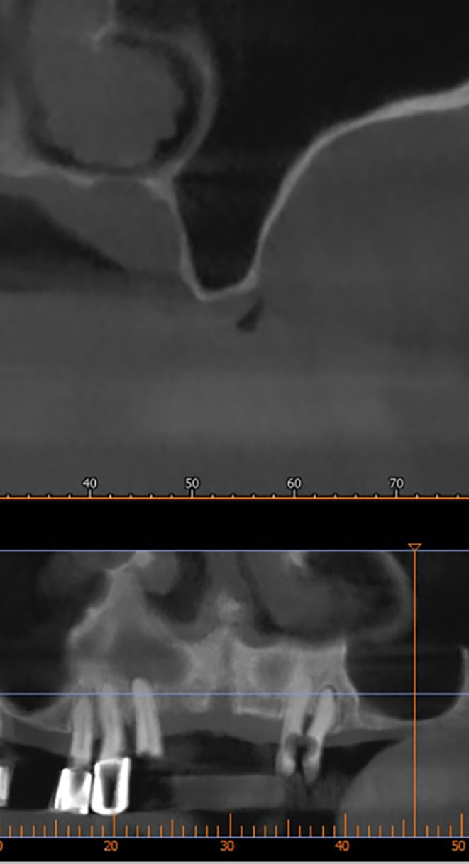 Figure 1
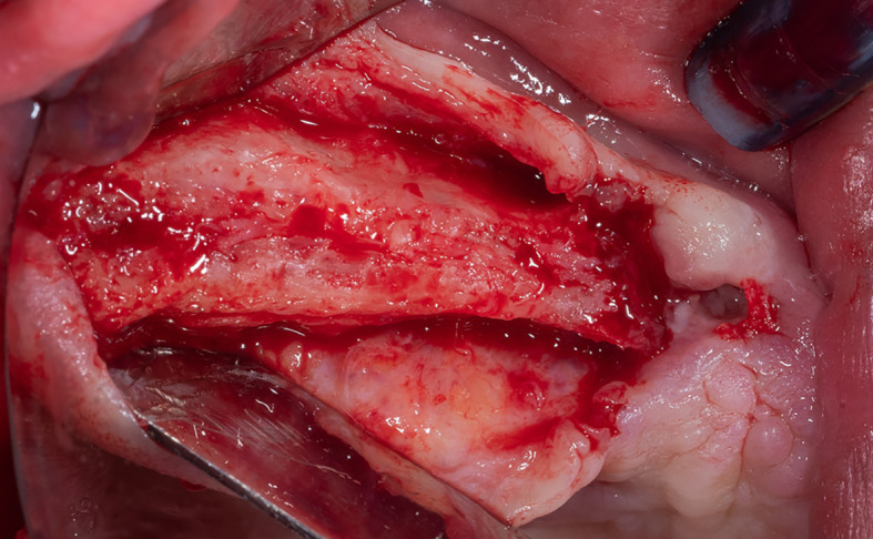 Figure 2
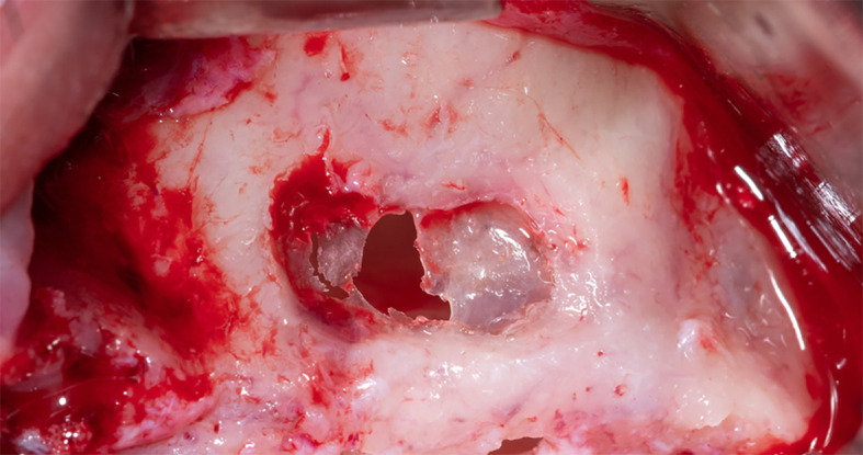 Figure 3
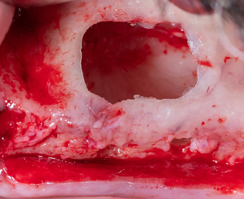 Figure 4
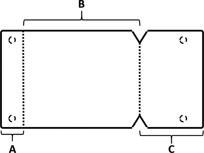 Figure 5
 Figure 6
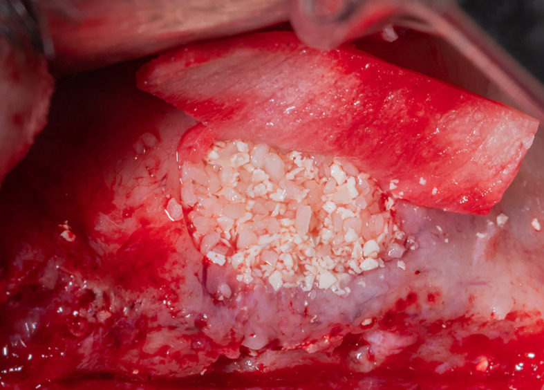 Figure 7
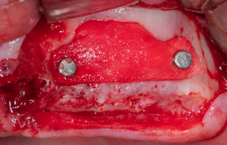 Figure 8
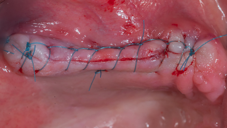 Figure 9
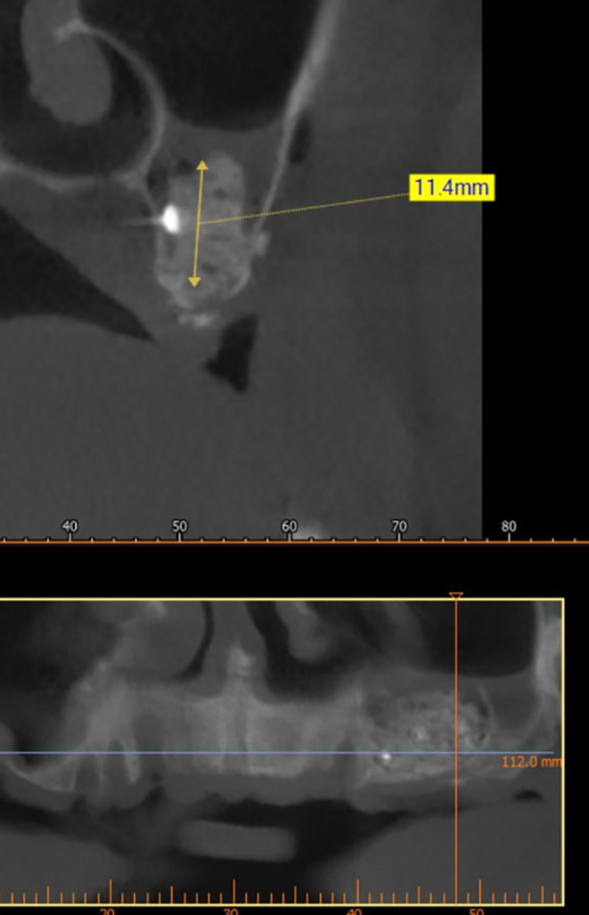 Figure 10
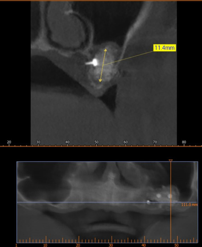 Figure 11
Peer Reviews
No public reviews on file for this paper yet. If you reviewed it on a platform where reviews are public (OpenReview, ICLR, NeurIPS, ICML), you can paste yours below so the community can read it here.
Videos
No videos yet. Explain this paper in a talk, walkthrough, or lecture? Add one.
Taxonomy
TopicsVoice and Speech Disorders · Facial Nerve Paralysis Treatment and Research · Temporomandibular Joint Disorders
1. Introduction
Dental implants are the best option to compensate for partial and complete tooth loss, especially in free edentulous ridge, and they also improve the quality of life for individuals. ^ 1 ^
However, they suffer from some limitations, such as important anatomical structures, which often have a lack of bone height, such as the maxillary sinus. ^ 2 ^ However, the development of many methods, such as the external sinus lift using the lateral approach, enabled us to perform dental implants when there is no bone opposite the maxillary sinus. ^ 2 ^
However, some limitations hindered the external sinus lift procedure, such as the thickness of the membrane, which may tear and hinder the completion of the external sinus lift process. ^ 3 ^Many grafting materials have been proved to enhance the antral area, such as autografts, ^ 2 ^ hydroxyapatite, ^ 3 ^ xenografts, ^ 4 ^ and various combinations of these. ^ 3 ^
Regardless of the chosen graft material, the sinus augmentation technique requires the elevation of the Schneiderian membrane and the insertion of the graft material into the area beneath the lifted membrane. ^ 5 ^
The most frequent sequela during sinus graft surgery is the tearing of the Schneiderian membrane depending on the thickness of the memebrane. ^ 6 ^
In case of a membrane perforation, the gap can be closed using a piece of resorbable collagen membrane or by suturing the Schneiderian membrane using a resorbable suture. ^ 7 ^
The Loma Linda Pouch technique was proposed by Proussaefs et al., ^ 8 ^ to create an artificial pouch from collagen membrane and the bony walls of the maxillary sinus and fill it with bone graft to solve the problem of Schneiderian membrane perforation.
Its modification was made by Das et al., ^ 9 ^ and this modification requires the membrane to be fixed at the palatal wall of the maxillary sinus and at the lateral window using sutures.
The present study shows a new development in this technique by modifying the design of the absorbable membrane and using bins to fix the membrane.
2. Case presentation
A non-smoker, medically free patient, aged 50 years, with need of dental implantation in the upper left area.
The patient participating in this study was informed of the details of the surgical procedure, the risks associated with it, and the alternative treatments. All his inquiries were answered and he signed the informed consent through which he agreed to enter the study and to the use of images of the surgical procedure in scientific articles.
After implant planning using the cone-beam computed tomography CBCT, it was found that the alveolar bone height corresponding to the maxillary sinus floor was (0.5-1 mm) ( Figure 1), which indicates an external sinus lift.
CBCT shows the thickness of the maxillary sinus floor.
Local anesthesia was performed by infiltration in the surgical area using 4% articaine with 1:80,000 adrenaline, a full-thickness gingival flap was lifted ( Figure 2), and a lateral window was opened in the maxillary sinus wall using Piezo electric device (PIEZOSURGERY®, Mectron, Italy) ( Figure 3).
Full-thickness gingival flap lifting.
The lateral window opening using Piezo electric device.
The sinus membrane was thin; therefore, it was torn when trying to open the lateral window and lift it, which prompted us to apply the pouch technique, which includes the following:
The membrane lifted from the inner sinus walls. Once the final intervention window is prepared ( Figure 4), we prepare the absorbable collagen membrane (Jason® membrane, Botiss, Germany) using the modified design performed by the researcher as it was used in this study for the first time ( Figure 5), where the membrane is measured and divided imaginarily into three parts as follows:
- A:4 mm wide, faces the palatal wall, and is fixed with it using pins.
- B: the same width as the maxillary sinus’s buccal palatal width and forms the roof of the artificially formed pouch.
- C:2 mm larger than the lateral window.
The final window after lifting schneiderian membrane.
The novel design of the collagen membrane.
Part A is determined and fixed to the palatal wall of the maxillary sinus using 2 pins (titan pin®, Botiss, Germany) and with a height equal to the future implants’ length + 2 mm ( Figure 6).
Fixing the collagen membrane to the palatal wall using pins.
After fixing the membrane to the palatal wall, the formed pouch is filled with a mixture of allograft (MYBONE, MSBIO, Korea) and xenograft (cerabone, Botiss, Germany) (30:70) ^ 10 ^ until it is filled ( Figure 7). Part B forms the roof of the formed pouch, compensating for the torn sinus membrane.
Filling the formed pouch with the bone graft mixture.
After that, the lateral window is closed with part C of the collagen membrane and fixed using pins medial and lateral to the window ( Figure 8), each pin is 2 mm away from the window border.
Fixing the collagen membrane using pins to close the lateral window.
Mechanical fixing of the membrane using pins and filling the formed pouch contributes to bone graft stability in the formed sinus and prevents the displacement of both the membrane and the bone graft mixture.
The gingival flap is then repositioned and sutured ( Figure 9). The patient was given postoperative instructions including not to rinse for 24 hours and to follow a soft diet during the first week after surgery. The patient was also given oral hygiene instructions.
The suture of the gingival flap.
An antibiotic (Amoxicillin 875 mg and Clavulanic acid 125 mg) was prescribed every 12 hours for 7 days and Diclofenac potassium 50 mg when needed, not to exceed 150 mg daily, in addition to chlorhexidine 0.12% mouthwash twice daily, 60 seconds for 14 days starting a day after surgery.
The surgical sutures were removed 14 days after surgery, and no sequelae occurred during the follow-up period that extended to nine months.
A CBCT scan was performed immediately after grafting and showed a direct bone gain of (11.4 mm) ( Figure 10) and 9 months after bone grafting where subsequent bone gain (11.4 mm) was found ( Figure 11).
CBCT after grafting immediately.
CBCT 9 months after grafting.
3. Discussion
Schneiderian membrane’s perforation is the most frequent sequela seen during sinus grafting. ^ 11 ^ Jensen et al., ^ 12 ^ report an occurrence of Schneiderian membrane perforation in 35% of cases, while others ^ 13 ^ report a 7% to 56% frequency when sinus grafting.
Several studies have advised the use of a resorbable collagen membrane to repair the perforated sinus membrane. ^ 14 ^
The aforementioned pouch technique may provide high protection and isolation of the graft material when conducting sinus grafting procedures. Furthermore, the creation of a pouch in the aforementioned technique presents the opportunity to cover the lateral access window with the collagen membrane employed to close the perforated area. This may cause increased isolation of the graft material. ^ 9 ^
Enhanced bone formation was observed in the grafted maxillary sinus when a non-resorbable barrier was applied to protect the graft material. However, other authors have suggested the resorbable collagen membrane use. ^ 15 ^
The Loma Linda pouch technique may provide enhanced isolation and protection of the graft material in sinus grafting procedures, particularly in cases where a perforation of the sinus membrane has occurred. ^ 8 ^
In the original proposal for the Loma Linda Pouch technique, ^ 8 ^ the collagen membrane is left unfixed and is stabilized by filling the pouch with bone graft, while in, ^ 9 ^ it is stabilized using sutures. This differs from the current study, which relied on a membrane design such that it is stabilized with bins while increasing the bone contact between the graft material and the bone walls.
However, a histological study is required to include histologic specimens that demonstrate bone formation within the sinus area.
4. Conclusion
Among the limitations of the current study, the novel design of the collagen membrane and its fixation with the bins led to greater stability of the bone graft and led to subsequent bone gain that enables dental implantation, but this technique requires a histological study to determine the nature of the bone formed.
Ethics approval and consent to participate
This study is approved by the ethical committee of Damascus University with number (DN-DN-020624-235). The study was registered on 5/16/2021 at Damascus University and the study start date was 9/21/2021. The patient participating in this study was informed of the details of the surgical procedure, the risks associated with it, and the alternative treatments. All his inquiries were answered and he signed the written informed consent through which he agreed to enter the study and to the use of images of the surgical procedure in scientific articles.
Consent to participate
The patient participating in this study was informed of the details of the surgical procedure, the risks associated with it, and the alternative treatments. All his inquiries were answered and he Written informed consent through which he agreed to enter the study and to the use of images of the surgical procedure in scientific articles.
The reference list from the paper itself. Each links out to its DOI / PubMed record.
- 1Pandey C Rokaya D Bhattarai BP : Contemporary concepts in osseointegration of dental implants: a review. Biomed. Res. Int. 2022;2022(1):6170452. 10.1155/2022/6170452 35747499 PMC 9213185 · doi ↗ · pubmed ↗
- 2Pogacian-Maier A-C : The Use of Allograft Bone in the Lateral Approach of Sinus Floor Elevation: A Systematic Review of Clinical Studies. Medicina. 2024;60(2):252. 10.3390/medicina 60020252 38399540 PMC 10890160 · doi ↗ · pubmed ↗
- 3Pesce P : Radiographic and histomorphometric evaluation of biomaterials used for lateral sinus augmentation: A systematic review on the effect of residual bone height and vertical graft size on new bone formation and graft shrinkage. J. Clin. Med. 2021;10(21):4996. 10.3390/jcm 10214996 34768518 PMC 8584826 · doi ↗ · pubmed ↗
- 4Correia F : Advantages of porcine xenograft over autograft in sinus lift: A randomised clinical trial. Materials. 2021;14(12):3439. 10.3390/ma 14123439 34205826 PMC 8234120 · doi ↗ · pubmed ↗
- 5Amid R : Effect of Schneiderian membrane thickening on the maxillary sinus augmentation and implantation outcomes: a systematic review. Journal of Maxillofacial and Oral Surgery. 2021;20:534–544. 10.1007/s 12663-021-01551-y 34776681 PMC 8554926 · doi ↗ · pubmed ↗
- 6Díaz-Olivares LA : Management of Schneiderian membrane perforations during maxillary sinus floor augmentation with lateral approach in relation to subsequent implant survival rates: a systematic review and meta-analysis. International Journal of Implant Dentistry. 2021;7:1–13. 10.1186/s 40729-021-00346-7 34250560 PMC 8273047 · doi ↗ · pubmed ↗
- 7Khoury F Schmidt C Jackowski J : The influence of suturing and or gluing of perforated Schneiderian membrane during sinuslift procedure on the outcome: a retrospective study. International Journal of Implant Dentistry. 2024;10(1):48. 10.1186/s 40729-024-00568-5 39496988 PMC 11534915 · doi ↗ · pubmed ↗
- 8Proussaefs P Lozada J : The "Loma Linda pouch": a technique for repairing the perforated sinus membrane. International Journal of Periodontics & Restorative Dentistry. 2003;23(6):593–597. 14703763 · pubmed ↗