Intraductal papillary mucinous neoplasm originating from a heterotopic pancreas within the stomach
Muyun Liu, Wei An, Jie Gao, Xingang Shi

Abstract
Genes, proteins, chemicals, diseases, species, mutations and cell lines named across the full text — each resolved to its canonical identifier and authoritative record.
Click any figure to enlarge with its caption.
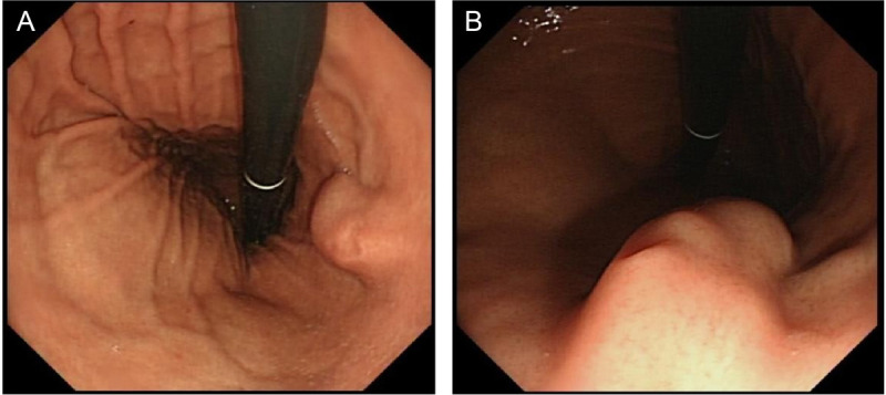 Figure 1
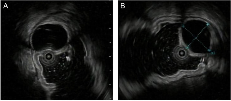 Figure 2
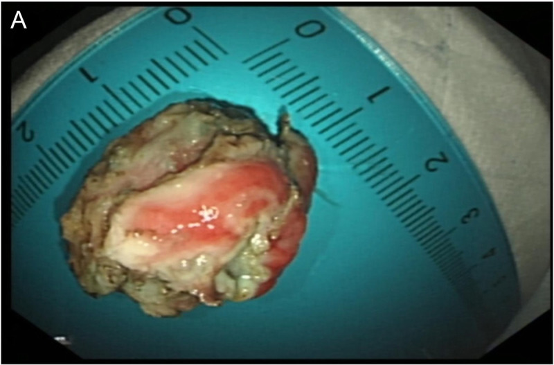 Figure 3
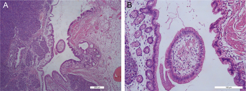 Figure 4
Peer Reviews
No public reviews on file for this paper yet. If you reviewed it on a platform where reviews are public (OpenReview, ICLR, NeurIPS, ICML), you can paste yours below so the community can read it here.
Videos
No videos yet. Explain this paper in a talk, walkthrough, or lecture? Add one.
Taxonomy
TopicsGastrointestinal disorders and treatments · Pancreatitis Pathology and Treatment · Pancreatic and Hepatic Oncology Research
A 32-year-old female patient was admitted due to the detection of a submucosal protrusion within the stomach in routine CT scan. The patient was asymptomatic without other history. Further gastroscopic examination showed that a hemispherical elevation measuring approximately 1.0 cm × 2.0 cm was identified on the posterior wall of the gastric body, with a visible depression on the surface under white-light observation [Figure 1A, B]. EUS revealed an oval mass sized 1.3 cm × 1.2 cm, protruding into and out of gastric lumen with clear boundaries, originating from the submucosal layer, with heterogeneous internal echoes, and local areas of medium to high echogenicity, including anechoic cystic spaces [Figure 2A, B]. Endoscopic submucosal dissection (ESD) was performed for diagnostic and therapeutic purposes. A tough nodular tumor measuring 2 cm × 1.5 cm × 1.5 cm was excised [Figure 3A]. Pathology revealed a gray-white, solid mass of medium consistency, and the pathological diagnosis was ectopic pancreas with intraductal papillary mucinous neoplasm (IPMN) (gastric type) formation accompanied with mild atypical hyperplasia [Figure 4A, B]. The patient experienced no discomfort postoperatively and discharged soon.
Ectopic pancreas typically presents as hypoechoic, isoechoic, or mixed echoic lesions under EUS. It can occur in any layer of the gastrointestinal wall, but most commonly in the submucosa, and can grow transmurally. The presence of duct-like structures is highly indicative for diagnosis.^[1]^ IPMN is a cystic lesion with malignant potential.^[2]^ Cases of ectopic pancreas complicated by IPMN are rare and only a few cases reported internationally.^[3–10]^ Among these cases, 3 developed malignancy. Therefore, early diagnosis is crucial for a favorable prognosis in such patients. Studies show that EUS has a sensitivity and specificity of 64% and 80%, respectively, for differentiating benign from malignant tumors, and it is superior to CT and MRI for lesions <2 cm in diameter.^[11]^ In our case, preoperative diagnosis of ectopic pancreas was challenging, and EUS-FNA offered a unique advantage.^[11]^ However, given the patient's young age, the patient opted for resection. Therefore, ESD was performed to remove the lesion following the guideline for the treatment of submucosal tumors (SMTs) within the gastrointestinal tract and revealed a favorable prognosis.
The reference list from the paper itself. Each links out to its DOI / PubMed record.
- 1Brun-Vergara ML Khoshpouri P Karp J Sailer A Pickhardt PJ. Heterotopic pancreatitis. Radiographics 2024;44(1):e 230167.38096108 10.1148/rg.230167 · doi ↗ · pubmed ↗
- 2Ohtsuka T Fernandez-Del Castillo C Furukawa T, . International evidence-based Kyoto guidelines for the management of intraductal papillary mucinous neoplasm of the pancreas. Pancreatology 2024;24(2):255–270.38182527 10.1016/j.pan.2023.12.009 · doi ↗ · pubmed ↗
- 3Huang JH Guo W Liu Z. Intraductal papillary mucinous neoplasm originating from a jejunal heterotopic pancreas: a case report. World J Clin Cases 2023;11(11):2496–2501.37123302 10.12998/wjcc.v 11.i 11.2496 PMC 10131005 · doi ↗ · pubmed ↗
- 4Pang Y Liu Y Liu Q Hou G. Intraductal papillary mucinous neoplasm arising from heterotopic pancreas in stomach: a case report and review of literature. Int J Surg Pathol 2023;31(5):708–713.35946106 10.1177/10668969221117990 · doi ↗ · pubmed ↗
- 5Patel N Berzin T. Intraductal papillary mucinous neoplasm arising in a heterotopic pancreas: a case report. Am J Gastroenterol 2010;105(11):2513–2514.21048696 10.1038/ajg.2010.298 · doi ↗ · pubmed ↗
- 6Cates JMM Williams TL Suriawinata AA. Intraductal papillary mucinous adenoma that arises from pancreatic heterotopia within a meckel diverticulum. Arch Pathol Lab Med 2005;129(3):e 67–e 69.15737052 10.5858/2005-129-e 67-IPMATA · doi ↗ · pubmed ↗
- 7Tsapralis D Charalabopoulos A Karamitopoulou E, . Pancreatic intraductal papillary mucinous neoplasm with concomitant heterotopic pancreatic cystic neoplasia of the stomach: a case report and review of the literature. Diagn Pathol 2010;5:4.20205774 10.1186/1746-1596-5-4PMC 2823681 · doi ↗ · pubmed ↗
- 8Okamoto H Fujishima F Ishida K, . Intraductal papillary mucinous neoplasm originating from a jejunal heterotopic pancreas: report of a case. Surg Today 2014;44(2):349–353.23325495 10.1007/s 00595-012-0486-0 · doi ↗ · pubmed ↗