A rare case of hepatic epithelioid hemangioendothelioma diagnosed by EUS-guided fine-needle biopsy (with videos)
Yating Wang, Beiyao Zhang, Dongqiang Zhao

Abstract
Genes, proteins, chemicals, diseases, species, mutations and cell lines named across the full text — each resolved to its canonical identifier and authoritative record.
Click any figure to enlarge with its caption.
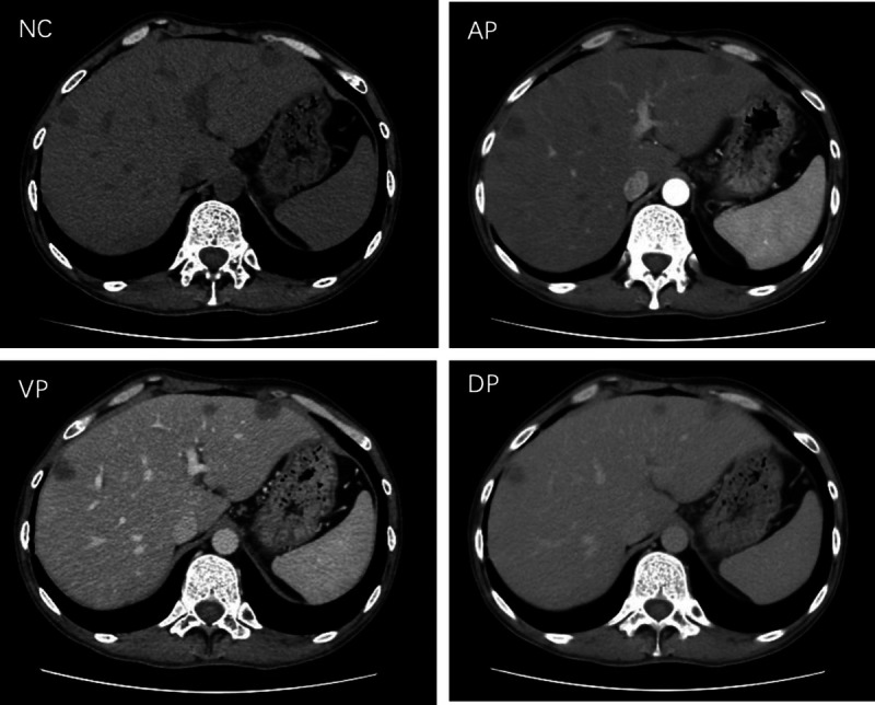 Figure 1
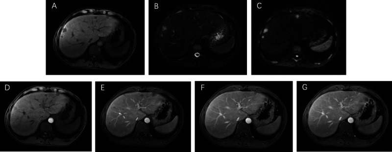 Figure 2
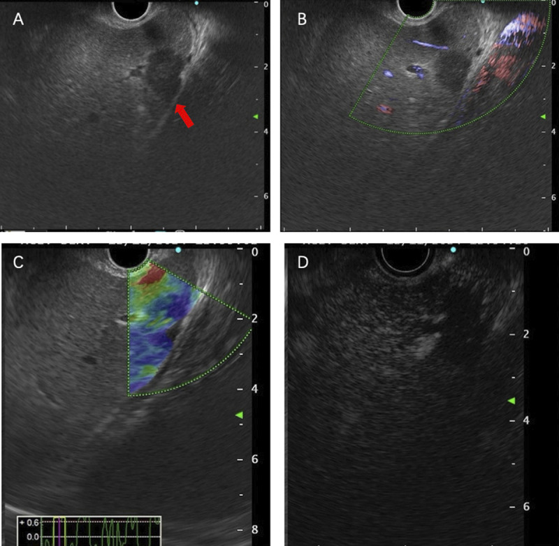 Figure 3
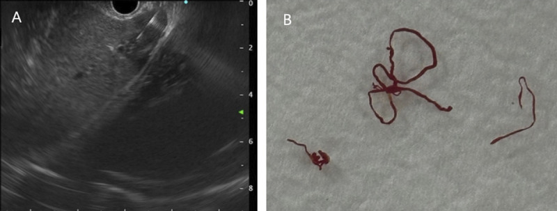 Figure 4
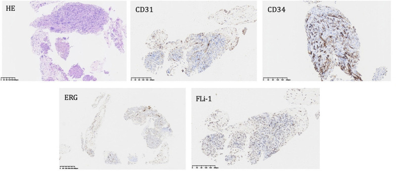 Figure 5
Peer Reviews
No public reviews on file for this paper yet. If you reviewed it on a platform where reviews are public (OpenReview, ICLR, NeurIPS, ICML), you can paste yours below so the community can read it here.
Videos
No videos yet. Explain this paper in a talk, walkthrough, or lecture? Add one.
Taxonomy
TopicsVascular Tumors and Angiosarcomas · Acute Myeloid Leukemia Research · Eosinophilic Disorders and Syndromes
A 35-year-old man was admitted to our department, with a 9-day history of jaundice. Preadmission liver function tests indicated hepatocellular damage and cholestasis, and abdominal ultrasound revealed multiple heterogeneous echoic masses in the liver. Tumor marker tests showed the following: alpha fetoprotein (AFP), 37.05 ng/mL; carcinoembryonic antigen (CEA), 4.47 ng/mL; carbonic anhydrase (CA)-125, 46.14 U/mL; and CA-199, 385.37 U/mL. Abdominal contrast-enhanced CT suggested multiple hypodense lesions in the liver [Figure 1]. Abdominal magnetic resonance imaging (MRI) revealed multiple intrahepatic lesions with abnormal signals [Figure 2A–B], some subcapsular, with hepatic capsular retraction. Diffusion-weighted imaging (DWI) showed restricted diffusion with pronounced hyperintensity [Figure 2C]. On contrast-enhanced imaging, some lesions showed peripheral ring enhancement in the arterial phase, followed by centripetal enhancement during the venous and delayed phases, with prominent enhancement in the late delayed phase [Figure 2D–G]. These findings are highly suggestive of hepatic epithelioid hemangioendothelioma (HEHE). Given the location of the lesions, EUS-guided fine-needle biopsy (EUS-FNB) was performed for definitive diagnosis. EUS (GF-UCT260, Olympus) revealed multiple quasi-circular hypoechoic lesions in the left lobe of the liver, most at the liver margin, measuring approximately 15 mm × 12 mm, with well-defined borders and heterogeneous internal echoes [Figure 3A, Video 1]. Color Doppler flow imaging showed no significant blood flow signals within the lesions [Figure 3B]. Elastography displayed blue-green coloration [Figure 3C], and contrast-enhanced EUS imaging revealed weak enhancement [Figure 3D]. A 22-gauge FNB needle (EUS-22-1-N, Micro-Tech) was used to access the liver lesions via the stomach, with 2 passes made [Figure 4, Video 2]. No complications occurred post-biopsy. Immunohistochemistry showed positivity for CD31, CD34, cytokeratin pan (CKpan), erythroblastosis transformation-specific regulated gene (ERG), and friend leukemia virus integration 1 (FLi-1), with a Ki-67 index of 5% [Figure 5]. The final pathological diagnosis was HEHE. Given the unresectable hepatic lesions without distant metastases, liver transplantation was recommended.
Supplementary Videos
Video 1: EUS imaging showed the location and characteristics of the hepatic lesions in the left lobe of the liver; Video 2: EUS-FNB procedure with a 22-gauge FNB needle. Videos are only available at the official website of the journal (www.eusjournal.com).
HEHE is a rare neoplasm of vascular origin with a variable course.^[1]^ Its progression ranges from long-term survival without treatment to rapid deterioration and fatal outcomes. Clinically, it is often an incidental finding, with symptoms ranging from nonspecific signs to advanced liver failure.^[2]^ The definitive diagnosis of HEHE relies on histopathology, and this case underscores the critical role of EUS-FNB as a crucial tool for obtaining definitive tissue samples.
Acknowledgments
None.
Source of Funding
None.
Ethical Approval
None declared.
Informed Consent
We obtained written informed consent from the patient to publish this article.
Conflict of Interest
The authors declare no conflict of interest for this article.
Author Contributions
Yating Wang reviewed the literature and contributed to manuscript writing and video producing, Beiyao Zhang cared for the patient and prepared the figure. Dongqiang Zhao was the operator who performed the EUS-FNB and was responsible for the conceptualization and supervision. All authors issued final approval for the version to be submitted.
The reference list from the paper itself. Each links out to its DOI / PubMed record.
- 1Sardaro A Bardoscia L Petruzzelli MF Portaluri M. Epithelioid hemangioendothelioma: an overview and update on a rare vascular tumor. Oncol Rev 2014;8:259.25992243 10.4081/oncol.2014.259PMC 4419652 · doi ↗ · pubmed ↗
- 2Studer LL Selby DM. Hepatic epithelioid hemangioendothelioma. Arch Pathol Lab Med 2018;142:263–267.29372848 10.5858/arpa.2016-0171-RS · doi ↗ · pubmed ↗