Pointing the trident in the right direction: recognizing spinal neurosarcoidosis through a specific MRI pattern
Alena Khalil, Kevin J. Abrams, Márcio Luís Duarte, Leonardo Furtado Freitas

Abstract
Genes, proteins, chemicals, diseases, species, mutations and cell lines named across the full text — each resolved to its canonical identifier and authoritative record.
Click any figure to enlarge with its caption.
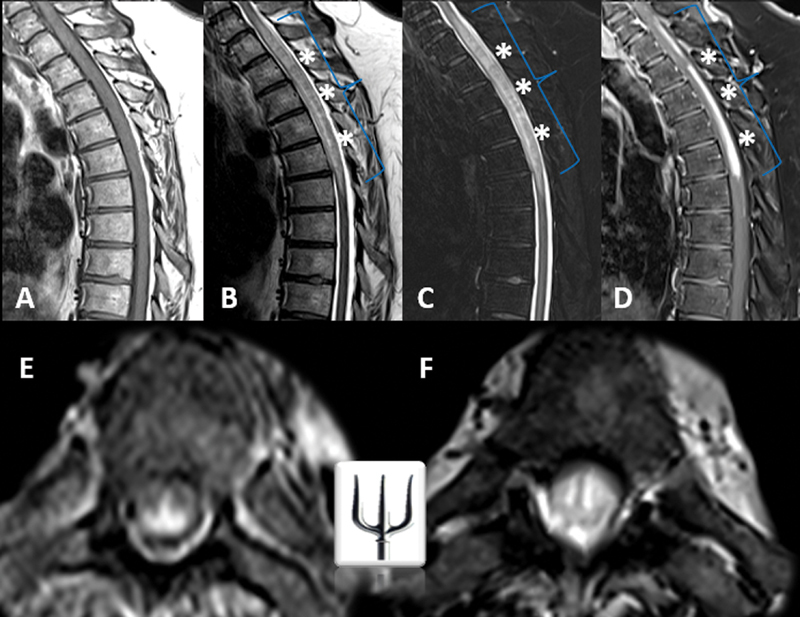 Figure 1
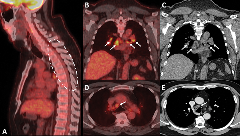 Figure 2
Peer Reviews
No public reviews on file for this paper yet. If you reviewed it on a platform where reviews are public (OpenReview, ICLR, NeurIPS, ICML), you can paste yours below so the community can read it here.
Videos
No videos yet. Explain this paper in a talk, walkthrough, or lecture? Add one.
Taxonomy
TopicsSarcoidosis and Beryllium Toxicity Research · Infectious Diseases and Tuberculosis · Medical Imaging Techniques and Applications
We herein report the case of a 51-year-old male presenting with progressive neurological symptoms, including numbness, tingling below the umbilicus, urinary difficulty, constipation, and weakness. Following a recent coronavirus disease 2019 (COVID-19) vaccination, magnetic resonance imaging (MRI) ( Figure 1 ) revealed longitudinally extensive transverse myelitis (LETM) with a trident-shaped pattern on axial sequences, a hallmark of spinal neurosarcoidosis. 1 2 3 Positron emission tomography-computed tomography ( Figure 2 ) demonstrated multiple hypermetabolic hilar and mediastinal lymphadenopathies, further supporting this diagnosis, particularly given the possibility of false-positive aquaporin-4-immunoglobulin G (AQP4-IgG) enzyme-linked immunosorbent assay (ELISA) results. 4 Early recognition of the trident sign enabled the prompt initiation of corticosteroids and immunosuppressive therapy, highlighting the diagnostic utility of this specific MRI pattern in an appropriate clinical scenario, with primary spinal cord lymphoma as a differential diagnosis. 5
Magnetic resonance imaging of the thoracic spine in sagittal ( A–D ) and axial views ( E–F ). Evidence of longitudinally extensive transverse myelitis at the cervicothoracic junction is observed, associated with significant edema and spinal cord swelling (blue braces). Notably, a contrast-enhancing component (white asterisks) in the posterior column and central regions, extending toward the pial/subpial surface and central ependymal canal, demonstrates T2-weighted/ short tau inversion recovery (STIR) hyposignal and exhibits a “trident-shaped head” appearance on axial images. The dorsal enhancement likely results from granulomatous inflammation spreading via perivascular pathways and may be influenced by meningeal lymphatic drainage and increased vascular permeability, predisposing this region to inflammation.
Positron emission tomography-computed tomography scan with fluorodeoxyglucose (FDG) in sagittal ( A ), coronal ( B–C ), and axial ( D–E ) views. No significant FDG uptake was observed in the cervicothoracic spinal cord lesion. However, multiple mediastinal, hilar, and precarinal lymphadenopathies demonstrated high radiotracer uptake, suggesting a granulomatous inflammatory process.
The reference list from the paper itself. Each links out to its DOI / PubMed record.
- 1Gibbons E Whittam D Jacob A Huda S Images of the month 1: Trident sign and neurosarcoidosis Clin Med (Lond)20212106 e 667e 66810.7861/clinmed.2021-059634862230 PMC 8806289 · doi ↗ · pubmed ↗
- 2Zalewski N L Krecke K N Weinshenker B G Aksamit A J Conway B L Mc Keon A Flanagan E P Central canal enhancement and the trident sign in spinal cord sarcoidosis Neurology 2016870774374410.1212/WNL.000000000000299227527540 · doi ↗ · pubmed ↗
- 3Beitollahi A Berry H Gulotta P Morales R Milburn J Trident Sign: The Key Magnetic Resonance Imaging Finding Distinguishing Spinal Cord Sarcoidosis From Multiple Sclerosis and Seropositive Neuromyelitis Optica Spectrum Disorder Ochsner J 2024240429830210.31486/toj.24.002739720826 PMC 11666104 · doi ↗ · pubmed ↗
- 4Jolliffe E A Keegan B M Flanagan E P Trident sign trumps Aquaporin-4-Ig G ELISA in diagnostic value in a case of longitudinally extensive transverse myelitis Mult Scler Relat Disord 2018237810.1016/j.msard.2018.04.01229709797 · doi ↗ · pubmed ↗
- 5Griffin K J Toledano M Flanagan E P Mustafa R“Trident Sign” in Primary CNS B-Cell Spinal Cord Lymphoma Neurology 20231011985785810.1212/WNL.000000000020767137648533 PMC 10662997 · doi ↗ · pubmed ↗