Selective Detection of Fungal and Bacterial Glycans with Galactofuranose (Galf) Residues by Surface-Enhanced Raman Scattering and Machine Learning Methods
Julia Yu. Zvyagina, Robert R. Safiullin, Irina A. Boginskaya, Ekaterina A. Slipchenko, Konstantin N. Afanas‘ev, Marina V. Sedova, Vadim B. Krylov, Dmitry V. Yashunsky, Dmitry A. Argunov, Nikolay E. Nifantiev, Ilya A. Ryzhikov, Alexander M. Merzlikin, Andrey N. Lagarkov

TL;DR
This study uses SERS and machine learning to detect specific sugar structures in fungi and bacteria that are absent in humans, offering a potential diagnostic tool for infections.
Contribution
The first use of SERS and machine learning to distinguish glycans containing galactofuranose (Galf) residues from fungal and bacterial sources.
Findings
SERS combined with PCA, CIE, and logistic regression successfully identified Galf-containing glycans.
Machine learning models reliably distinguished glycan structures with and without Galf residues.
The approach complements traditional methods by highlighting key spectral features linked to Galf presence.
Abstract
Specific monosaccharide residue, β-D-galactofuranose (Galf) featuring a five-membered ring structure, is found in the glycans of fungi and bacteria, but is normally absent in healthy mammals and humans. In this study, synthetic oligosaccharides mimicking bacterial and fungal glycans were investigated by SERS (Surface-Enhanced Raman Scattering) techniques for the first time to distinguish between different types of glycan chains. SERS spectra of oligosaccharides related to fungal α-(1→2)-mannan, β-(1→3)-glucan, β-(1→6)-glucan, galactomannan of Aspergillus, galactan I of Klebsiella pneumoniae, and diheteroglycan of Enterococcus faecalis were measured. To analyze the spectra, a number of machine learning methods were used that complemented each other: principal component analysis (PCA), confidence interval estimation (CIE), and logistic regression with L1 regularization. Each of the…
Genes, proteins, chemicals, diseases, species, mutations and cell lines named across the full text — each resolved to its canonical identifier and authoritative record.
Click any figure to enlarge with its caption.
 Figure 1
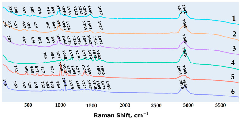 Figure 2
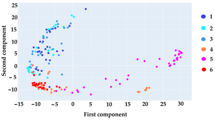 Figure 3
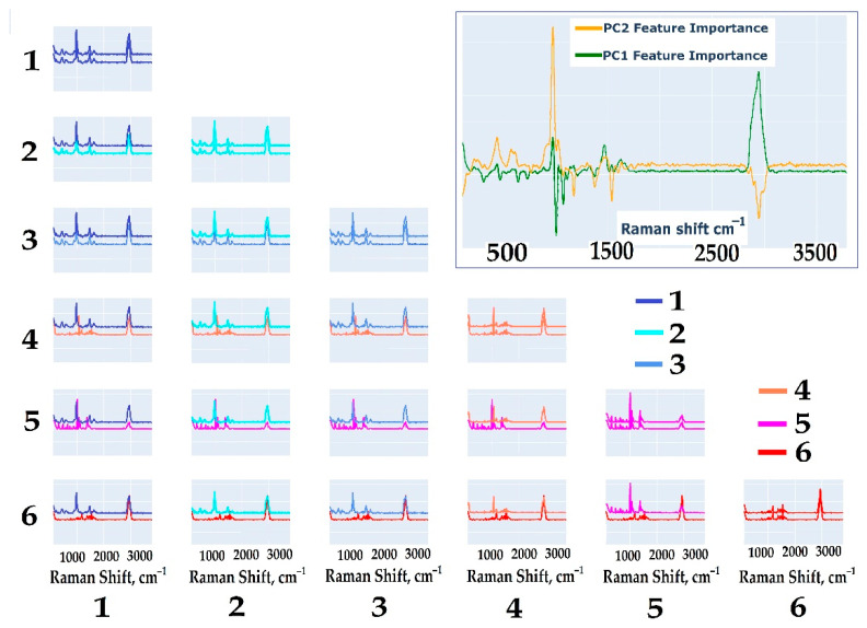 Figure 4
 Figure 5
 Figure 6
 Figure 7
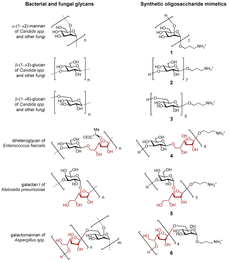 Figure 8
Peer Reviews
No public reviews on file for this paper yet. If you reviewed it on a platform where reviews are public (OpenReview, ICLR, NeurIPS, ICML), you can paste yours below so the community can read it here.
Videos
No videos yet. Explain this paper in a talk, walkthrough, or lecture? Add one.
Taxonomy
TopicsSpectroscopy Techniques in Biomedical and Chemical Research · Probiotics and Fermented Foods · Spectroscopy and Chemometric Analyses