Computer-Aided Detection (CADe) of Small Metastatic Prostate Cancer Lesions on 3D PSMA PET Volumes Using Multi-Angle Maximum Intensity Projections
Amirhosein Toosi, Sara Harsini, Ghasemali Divband, François Bénard, Carlos F. Uribe, Felipe Oviedo, Rahul Dodhia, William B. Weeks, Juan M. Lavista Ferres, Arman Rahmim

TL;DR
This paper introduces a new automated system to detect small prostate cancer metastases in PET scans using advanced AI techniques, aiming to improve diagnosis and reduce workload for doctors.
Contribution
The novel approach combines multi-angle projections with 2D object detection models to efficiently detect small metastatic lesions in 3D PSMA-PET volumes.
Findings
The FreeAnchor model achieved an F1-score of 0.69 and a recall of 0.74 for lesion detection.
The system showed strong recall rates for local relapses (0.82) and bone metastases (0.80).
The method outperformed several 3D methods in efficiency while maintaining high accuracy.
Abstract
We aimed to develop an automated computer-aided detection (CADe) system to help doctors detect small metastatic prostate cancer (PCa) lesions more efficiently, ultimately acting as a “second reader” to improve diagnosis and reduce workload in cancer care. Our method used multi-angle Maximum Intensity Projections (MA-MIPs) and explored state-of-the-art (SOTA) object detection AI algorithms. We evaluated 16 SOTA models across four categories. The system identified lesions in 2D images and then mapped them back into 3D space. A fine-tuned segmentation model further refined the results. Our best model, FreeAnchor, achieved a stronger detection performance. It was more efficient than many 3D methods while maintaining high accuracy, and it performed especially well for local relapses and bone metastases. Objectives: We aimed to develop and evaluate a novel computer-aided detection (CADe)…
Genes, proteins, chemicals, diseases, species, mutations and cell lines named across the full text — each resolved to its canonical identifier and authoritative record.
Click any figure to enlarge with its caption.
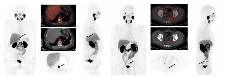 Figure 1
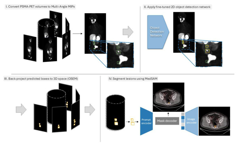 Figure 2
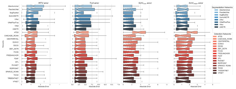 Figure 3
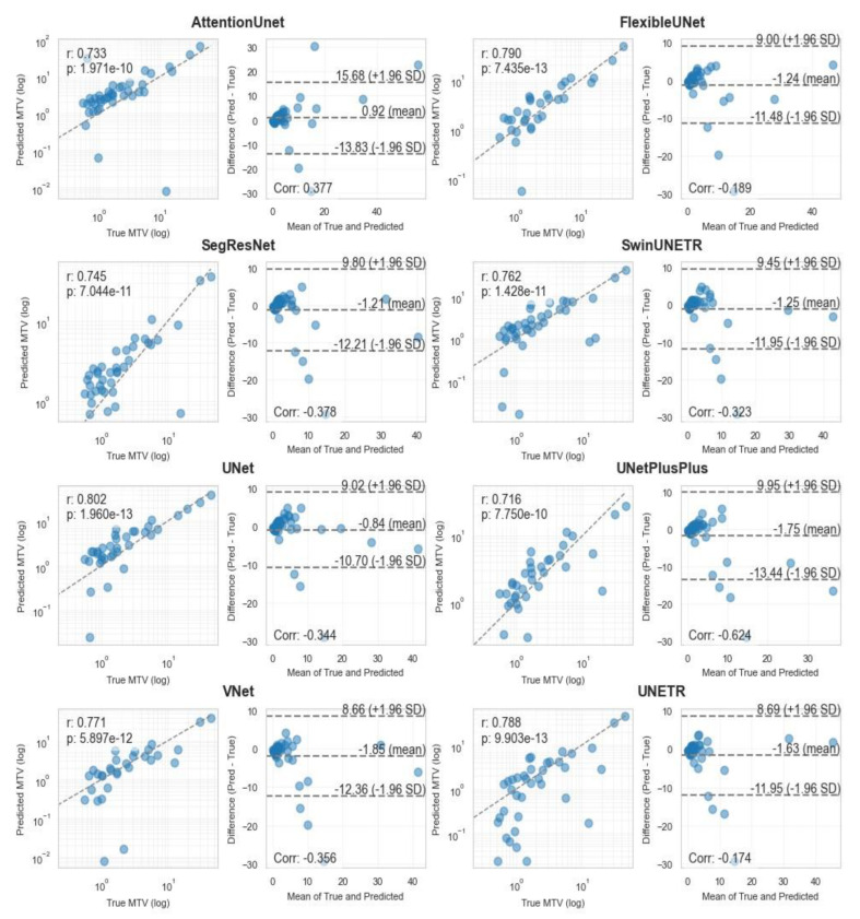 Figure 4
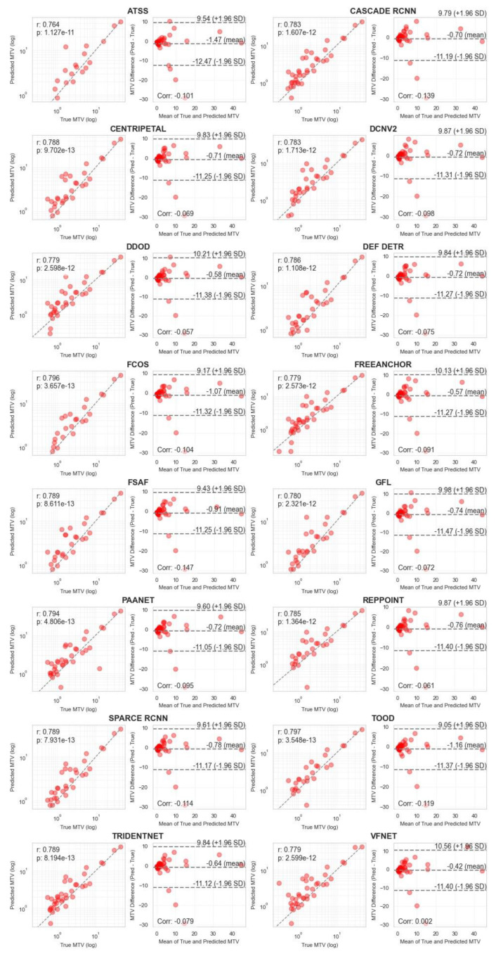 Figure 5
Peer Reviews
No public reviews on file for this paper yet. If you reviewed it on a platform where reviews are public (OpenReview, ICLR, NeurIPS, ICML), you can paste yours below so the community can read it here.
Videos
No videos yet. Explain this paper in a talk, walkthrough, or lecture? Add one.
Taxonomy
TopicsAdvanced Neural Network Applications · Radiomics and Machine Learning in Medical Imaging · AI in cancer detection