Thoracic CT Angiographies in Children Using Automated Power Injection with Bolus Tracking Versus Manual Contrast Injection: Analysis of Contrast Enhancement, Image Quality and Radiation Exposure
Jochen Pfeifer, Deborah Driulini, Katrin Altmeyer, Gudrun Wagenpfeil, Martin Poryo, Christian Giebels, Arno Bücker, Alexander Massmann, Hashim Abdul-Khaliq, Peter Fries

TL;DR
This study compares automated and manual contrast injection methods in pediatric CT angiography, finding automated methods provide better image quality without significantly increasing radiation exposure.
Contribution
Demonstrates that automated power injection with bolus tracking improves image quality in pediatric thoracic CTA compared to manual injection.
Findings
Automated injection resulted in significantly higher attenuation and contrast-to-noise ratios in key cardiac structures.
Image quality was rated significantly better with automated injection (Likert 3–4) compared to manual (Likert 2–3).
Radiation exposure was not significantly different between the two methods.
Abstract
Objectives: The purpose of this study was to analyze image quality and radiation exposure of thoracic computed tomography angiography (CTA) in children with congenital heart diseases (CHDs) using either manual contrast medium (CM) injection or automated power injectors with bolus tracking. Methods: A total of 137 thoracic CTAs of 120 consecutive pediatric patients were included in this retrospective study. We analyzed the method of CM administration (power injection with bolus tracking (PI) or manual injection (MI)), injection routes, volumes and flow rates of CM. For the evaluation of objective image quality, attenuation values in the heart chambers and great thoracic vessels were determined by region-of-interest (ROI) analysis and signal-to-noise (SNR) and contrast-to-noise (CNR) ratios calculated thereof. Visual image quality was assessed by two blinded readers (four-point…
Genes, proteins, chemicals, diseases, species, mutations and cell lines named across the full text — each resolved to its canonical identifier and authoritative record.
Click any figure to enlarge with its caption.
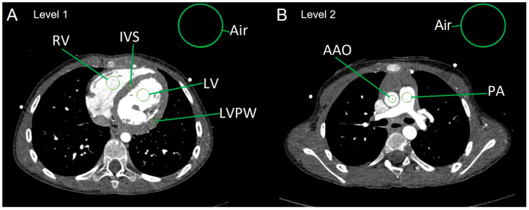 Figure 1
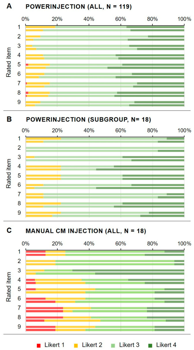 Figure 2
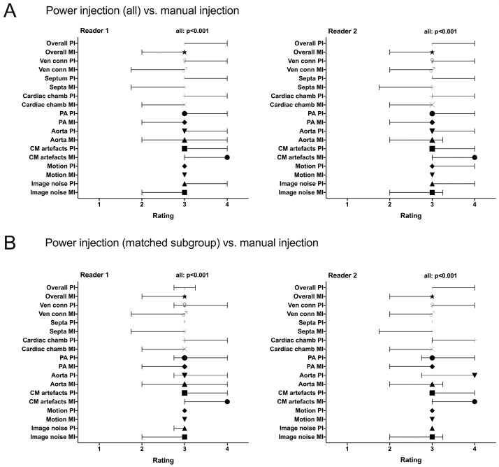 Figure 3
 Figure 4
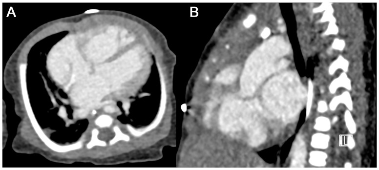 Figure 5
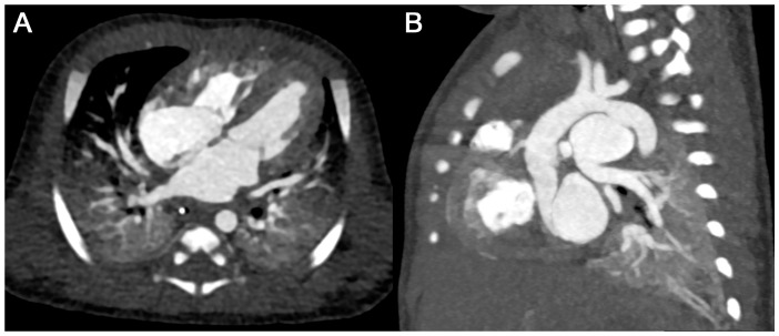 Figure 6
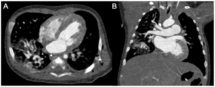 Figure 7
Peer Reviews
No public reviews on file for this paper yet. If you reviewed it on a platform where reviews are public (OpenReview, ICLR, NeurIPS, ICML), you can paste yours below so the community can read it here.
Videos
No videos yet. Explain this paper in a talk, walkthrough, or lecture? Add one.
Taxonomy
TopicsRadiation Dose and Imaging · Ultrasound in Clinical Applications · Advanced X-ray and CT Imaging