Cerebrolith in hydranencephaly
Mehmet Atalar

Abstract
Genes, proteins, chemicals, diseases, species, mutations and cell lines named across the full text — each resolved to its canonical identifier and authoritative record.
Click any figure to enlarge with its caption.
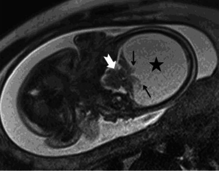 Figure 1
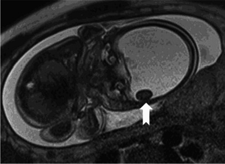 Figure 2
- —Sivas Cumhuriyet University
Peer Reviews
No public reviews on file for this paper yet. If you reviewed it on a platform where reviews are public (OpenReview, ICLR, NeurIPS, ICML), you can paste yours below so the community can read it here.
Videos
No videos yet. Explain this paper in a talk, walkthrough, or lecture? Add one.
Taxonomy
TopicsFetal and Pediatric Neurological Disorders · Head and Neck Surgical Oncology · Craniofacial Disorders and Treatments
A 34-year-old woman, gravida 2, parity 1, was referred from an external center at 26 weeks’ gestation with a preliminary diagnosis of hydrocephalus. The patient underwent fetal magnetic resonance imaging (MRI). Coronal T2-weighted (T2W) half-Fourier acquisition single-shot turbo spin-echo (HASTE) MR image (a) shows hydranencephaly (asterisk). Posterior fossa structures are intact (white arrow). Remnants of occipital parenchymal structures are also visible (black arrows). Coronal T2W HASTE MR image (b) shows a cerebrolith as a hypointense nodular mass adjacent to the inner table of the occipital bone (arrow). The cerebrolith associated with hydranencephaly may represent infarcted brain tissue, which is consistent with the etiology of hydranencephaly.