Spontaneous hip ankylosis in end-stage osteoarthritis: a rare case of complete bony fusion
Nikita Gangwani, Gurjeet Kaur

Abstract
Genes, proteins, chemicals, diseases, species, mutations and cell lines named across the full text — each resolved to its canonical identifier and authoritative record.
Click any figure to enlarge with its caption.
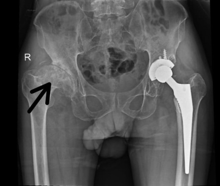 Figure 1
Peer Reviews
No public reviews on file for this paper yet. If you reviewed it on a platform where reviews are public (OpenReview, ICLR, NeurIPS, ICML), you can paste yours below so the community can read it here.
Videos
No videos yet. Explain this paper in a talk, walkthrough, or lecture? Add one.
Taxonomy
TopicsHip disorders and treatments · Bone and Joint Diseases · Bone health and treatments
Image in medicine
This case highlights a rare progression of hip osteoarthritis to spontaneous ankylosis, marked by unique clinical features. A 53-year-old male farmer presented with severe functional impairment due to complete loss of motion in his right hip, which progressed to painless ankylosis. Remarkably, the ankylosis occurred unilaterally without preceding trauma, inflammatory arthritis, or family history of joint disorders, contrasting with a well-functioning total hip replacement on the left side. Radiographs revealed total bony fusion of the right femoral head and acetabulum, with evident sclerosis and remodeling, while surrounding bone mineralization remained intact. The left hip arthroplasty components were well-positioned with no complications. This patient´s history, including a 50-pack-year smoking history and physically demanding occupation, likely contributed to accelerated joint degeneration and ankylosis. Muscle power around the right hip was graded at 2/5, with a 0° range of motion and notable iliopsoas and adductor tightness. Differential diagnoses such as post-traumatic ankylosis and ankylosing spondylitis were ruled out due to the absence of inflammatory markers or injury. This case is distinct as spontaneous ankylosis is a rare AVN outcome, particularly without surgical intervention. It underscores the need for early medical management of osteoarthritis and highlights the role of lifestyle and occupation in joint pathology. Its scientific significance lies in demonstrating the complex mechanisms of joint degeneration and the importance of personalized approaches to joint health.
the right hip shows complete loss of joint space with bony fusion between the femoral head and acetabulum, consistent with spontaneous ankylosis, sclerosis, and bone remodeling around the ankylosed joint; the left hip demonstrates well-positioned total hip arthroplasty components without evidence of loosening or hardware complications, and the remaining pelvic bones appear intact with normal mineralization