Massive cardiomegaly secondary to rheumatic heart disease
Gaurang Aurangabadkar, Sumer Choudhary

Abstract
Genes, proteins, chemicals, diseases, species, mutations and cell lines named across the full text — each resolved to its canonical identifier and authoritative record.
Click any figure to enlarge with its caption.
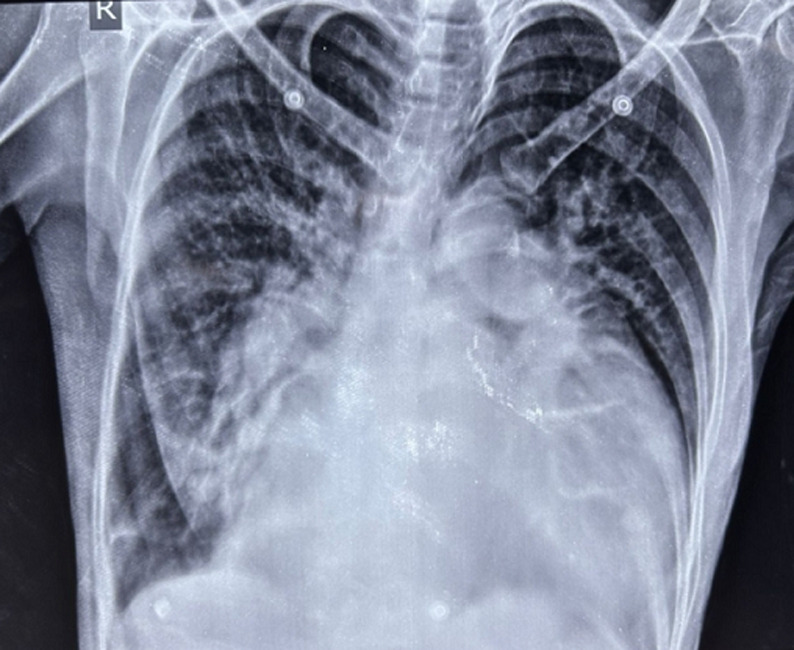 Figure 1
Peer Reviews
No public reviews on file for this paper yet. If you reviewed it on a platform where reviews are public (OpenReview, ICLR, NeurIPS, ICML), you can paste yours below so the community can read it here.
Videos
No videos yet. Explain this paper in a talk, walkthrough, or lecture? Add one.
Taxonomy
TopicsViral Infections and Immunology Research · Kawasaki Disease and Coronary Complications · Cardiac tumors and thrombi
Image in medicine
A 54-year-old female patient presented to the respiratory physician with chief complaints of dyspnea on exertion, dysphagia, and chest pain. The patient´s past medical history revealed a diagnosis of rheumatic heart disease, which was initially diagnosed 8 years back and a recent echocardiography report was suggestive of severe mitral stenosis with a left ventricular ejection fraction (LVEF) of 28%. An esophagoscopy was done given dysphagia which revealed no obvious abnormalities of the esophageal mucosa. A chest X-ray postero-anterior (PA) view was done which revealed the presence of a massive cardiomegaly with a cardiothoracic ratio of 0.80 (normal cardio-thoracic ratio <0.50). A cardiologist's opinion was taken and the patient was started on angiotensin-converting enzyme (ACE) inhibitors, oral furosemide (diuretic), and carvedilol (beta-blockers), along with regular follow-up. The patient was discharged with the same advice after 5 days of admission. Gross cardiomegaly is a rare complication of rheumatic heart disease, usually seen in patients with severe mitral stenosis, and occurs as a result of altered cardio-pulmonary hemodynamics arising as a result of valvular pathology. Such patients usually present with complaints of dyspnea and dysphagia arising as a result of the considerable enlargement of the cardiac dimensions. This clinical image aims to highlight this striking presentation of gross cardiomegaly that is seen to occupy more than 75% of the hemithorax in horizontal dimensions.
chest X-ray postero-anterior (PA) view demonstrating gross cardiomegaly in a patient with rheumatic heart disease