Endoscopic submucosal dissection for removal of a large hypopharyngeal lipoma with oval forceps traction
Tian-Xing Yuan, Ye-Han Zhou, Yu Bao, Rui Zhao

Abstract
Genes, proteins, chemicals, diseases, species, mutations and cell lines named across the full text — each resolved to its canonical identifier and authoritative record.
Click any figure to enlarge with its caption.
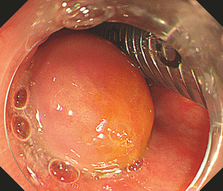 Fig. 1
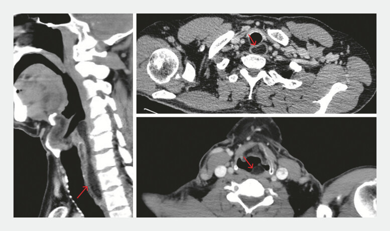 Fig. 2
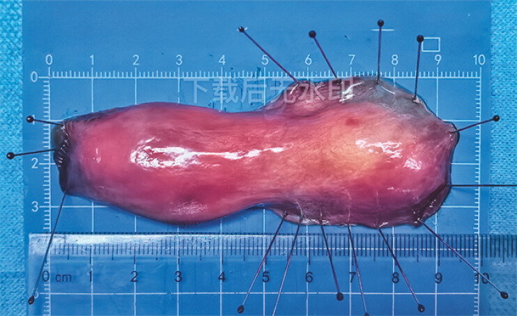 Fig. 3
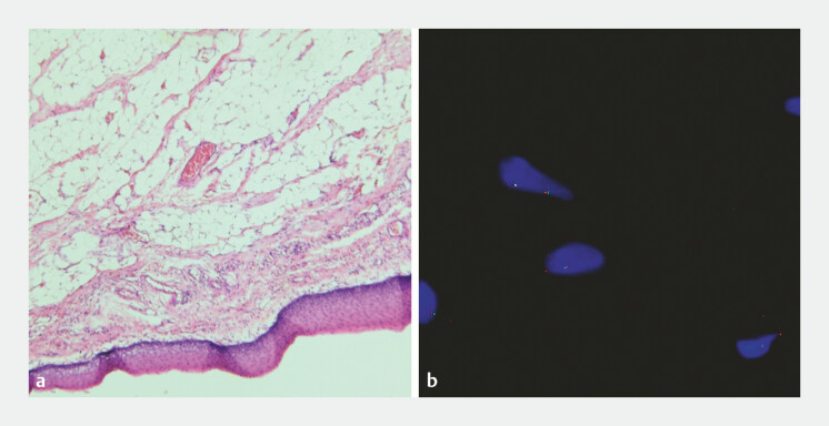 Fig. 4
- —The major technology application and demonstration project, Chengdu Science and Technology Bureau
Peer Reviews
No public reviews on file for this paper yet. If you reviewed it on a platform where reviews are public (OpenReview, ICLR, NeurIPS, ICML), you can paste yours below so the community can read it here.
Videos
No videos yet. Explain this paper in a talk, walkthrough, or lecture? Add one.
Taxonomy
TopicsTracheal and airway disorders · Tumors and Oncological Cases · Gastrointestinal disorders and treatments
A 59-year-old man presented with a mass in the hypopharynx found during routine screening. Endoscopic examination revealed a large submucosal swelling in the posterior hypopharynx and cervical esophagus, with the base located in the posterior hypopharyngeal wall ( Fig. 1 ). Contrast-enhanced CT showed a mass with fat density, which did not enhance ( Fig. 2 ), consistent with a lipoma.
Endoscopic findings. The base located in the posterior hypopharyngeal wall.
Contrast-enhanced CT showed a mass with fat density, without significant contrast enhancement.
The tumor was successfully removed ( Fig. 3 ) using endoscopic submucosal dissection (ESD) assisted by oval forceps ( Video 1 ). The oval forceps provided optimal traction, ensuring clear exposure of the broad tumor base, which facilitated its complete removal. Due to the broad base of the tumor, snare traction was relatively difficult to perform. Oval forceps, however, provided simpler operation and allowed for better visualization of the tumor base. The procedure took 57 minutes, with no major complications during or after the surgery.
The specimen measured 9.5 cm in length and 3.5 cm in width at its widest point.
Endoscopic submucosal dissection was performed to remove a large hypopharyngeal lipoma with oval forceps traction.Video 1
Histopathological analysis confirmed the diagnosis of submucosal lipoma. Fluorescence in situ hybridization (FISH) testing revealed no MDM2 gene amplification, excluding liposarcoma ( Fig. 4 ).
Histological analysis revealed a submucosal lipoma. a Hematoxylin and eosin-stained images (×50). b FISH testing revealed no MDM2 gene amplification. Abbreviation: FISH, fluorescence in situ hybridization.
Lipomas are common benign tumors but are rare in the hypopharynx 1 . To our knowledge, there have been reports using snare traction-assisted ESD to remove hypopharyngeal tumors 2 , but the use of oval forceps for ESD in large hypopharyngeal lipomas has not been previously described.
This case highlights the feasibility, safety, and effectiveness of oval forceps-assisted ESD in treating large hypopharyngeal lipomas, particularly those with a broad base. This method offers a practical, minimally invasive option for the endoscopic management of these rare tumors.
Endoscopy_UCTN_Code_CCL_1AB_2AB
Endoscopy_UCTN_Code_TTT_1AO_2AG_3AD
The reference list from the paper itself. Each links out to its DOI / PubMed record.
- 1Eo TS Shin HA Kie JH Pedunculated Fibrolipoma of the Hypopharynx: A Case Report J Korean Soc Laryngol Phoniatr Logop 202233115118
- 2Liu L Miao F Guo HM Endoscopic Submucosal Dissection of the Angiolipoma at Hypopharynx-Esophageal Introitus Gastroenterol Res Pract 202020201710.1155/2020/3581267 PMC 704250032148476 · doi ↗ · pubmed ↗