Dysphagia and thickened esophageal wall: the application of ultra-slim gastroscope in the diagnosis and treatment of phlegmonous esophagitis
Guangzhao Li, Huikai Li, Xiuxue Feng, Fen Deng

Abstract
Genes, proteins, chemicals, diseases, species, mutations and cell lines named across the full text — each resolved to its canonical identifier and authoritative record.
Click any figure to enlarge with its caption.
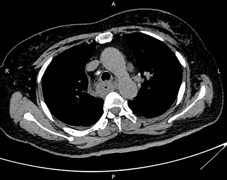 Fig. 1
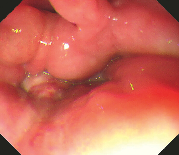 Fig. 2
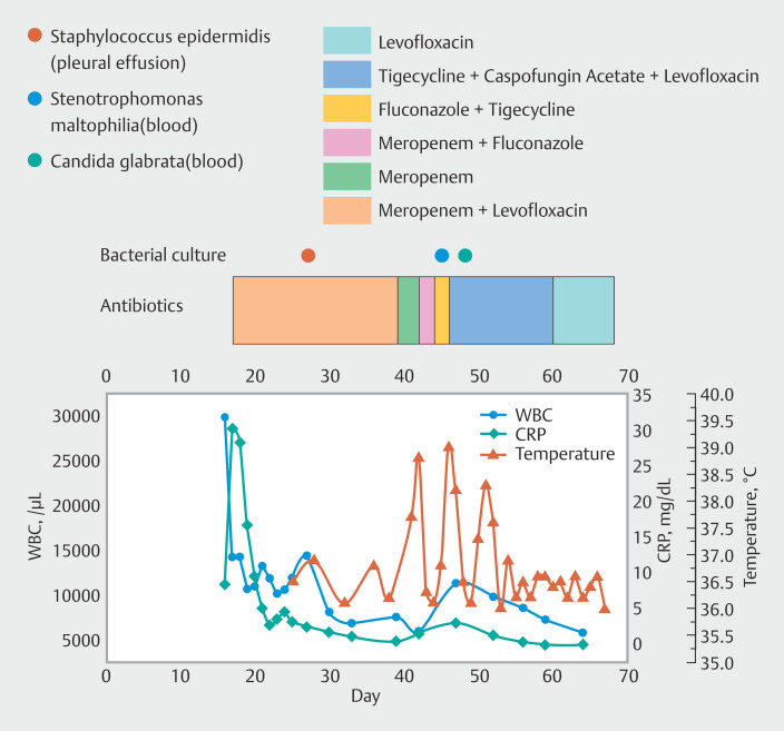 Fig. 3
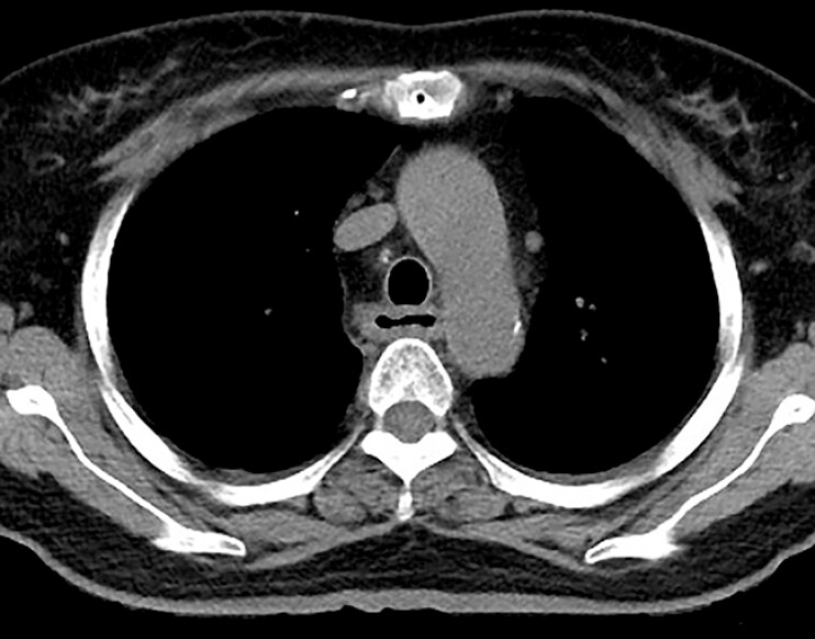 Fig. 4
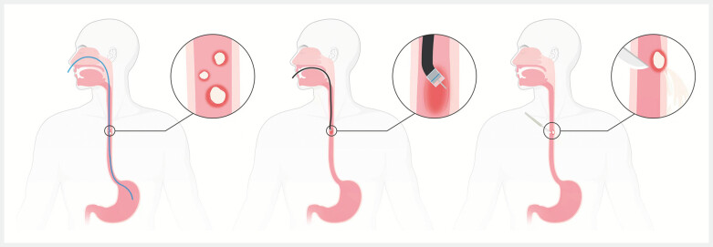 Fig. 5
- —National Key Research and Development Program of China10.13039/501100012166
Peer Reviews
No public reviews on file for this paper yet. If you reviewed it on a platform where reviews are public (OpenReview, ICLR, NeurIPS, ICML), you can paste yours below so the community can read it here.
Videos
No videos yet. Explain this paper in a talk, walkthrough, or lecture? Add one.
Taxonomy
TopicsGastroesophageal reflux and treatments · Esophageal and GI Pathology · Abdominal vascular conditions and treatments
A 61-year-old woman presented with persistent sore throat for 17 days and progressively worsening dysphagia for 13 days. Computed tomography (CT) of the chest showed a thickened wall of the entire esophagus and blurring of the peri-esophageal fat space ( Fig. 1 ). Standard gastroscope could not be passed through the esophageal entrance because of significant pharyngeal stenosis and edema, and pus oozing was seen at the entrance of the esophagus ( Fig. 2 ). Pharyngeal infection was considered. After intravenous antibiotics ( Fig. 3 ), infection was controlled but dysphagia remained without any sign of relief. On day 50, an ultra-slim gastroscope revealed three mucosal defects with a diameter of 4–10 mm in the esophagus 18–23 cm from the incisors. The endoscope was entered through the largest mucosal defect into the submucosal layer and white thin pus was found within several submucosal cavities, which formed after self-absorption of pus and indicated phlegmonous esophagitis ( Video 1 ). Enteral nutrition is performed through an endoscopic indwelling gastric tube. On day 55, chest CT showed a significant reduction of esophageal wall thickening ( Fig. 4 ). On day 63, repeat gastroscopy showed two of the three previous mucosal defects healed and one remained there with a size of 5 mm, which was closed by two metal clips. The patient then started eating orally and was discharged on day 70. Phlegmonous esophagitis is rare and there is no standard treatment for phlegmonous esophagitis 1 . Available treatment options include infection control with antibiotics, endoscopic incision 2 3 4 5 , or surgery ( Fig. 5 ). We report the first video of complete access into the abscess cavities of spontaneously ruptured phlegmonous esophagitis, which was achieved with a favorable therapeutic outcome by endoscopic placement of a gastric tube under direct visualization. We believe that gastric tube placement, rather than endoscopic incision or surgery, can result in good outcomes and enable early enteral nutrition in phlegmonous esophagitis with primary spontaneous rupture.
Endoscopy_UCTN_Code_CCL_1AB_2AC
Computed tomography of the chest showed a thickened wall of the entire esophagus and blurring of the peri-esophageal fat space.
Standard gastroscope could not be passed through the esophageal entrance because of significant pharyngeal stenosis and edema, and pus oozing was seen at the entrance of the esophagus.
Patient’s bacterial culture results, history of antibiotic therapy, laboratory results, and temperature changes since the patientʼs presentation in the clinic.
On day 55, computed tomography of the chest showed a significant reduction of esophageal wall thickening.
Available treatment options include infection control with antibiotics, endoscopic incision, or surgery. Created in BioRender. Li, G. (2025) https://BioRender.com/t39f227 . [rerif].
The application of ultra-slim gastroscope in the diagnosis and treatment of phlegmonous esophagitis.Video 1
The reference list from the paper itself. Each links out to its DOI / PubMed record.
- 1Jin DH Woo W Lee J Optimal Management of Patients with Phlegmonous Esophagitis: A Systematic Review and Meta-Analysis J Clin Med 202312714710.3390/jcm 1222714738002759 PMC 10672419 · doi ↗ · pubmed ↗
- 2Onana Ndong P Piche T Vanbiervliet G Comprehensive endoscopic management of recurrent esophageal wall abscess revealing concomitant eosinophilic esophagitis Endoscopy 202456 E 510E 51110.1055/a-2325-277038866058 PMC 11168795 · doi ↗ · pubmed ↗
- 3Saito Y Asami M Miki A Deep Neck Infection Complicated by Phlegmonous Esophagitis and Mediastinitis Ann Thorac Surg 2021111 e 403e 40610.1016/j.athoracsur.2020.08.10133232726 · doi ↗ · pubmed ↗
- 4Kim JW Ahn HY Kim GH Endoscopic Intraluminal Drainage: An Alternative Treatment for Phlegmonous Esophagitis Korean J Thorac Cardiovasc Surg 20195216516910.5090/kjtcs.2019.52.3.16531236377 PMC 6559182 · doi ↗ · pubmed ↗
- 5Tonouchi A Kuwabara S Furukawa K Phlegmonous esophagitis treated by endoscopic drainage Esophagus 20171418318710.1007/s 10388-016-0562-4 · doi ↗