Forward-viewing echoendoscope-guided pancreaticojejunostomy for post-pancreaticoduodenectomy stricture
Toru Kaneko, Mitsuhiro Kida, Takahiro Kurosu, Yutaro Saito, Shiori Koyama, Tomohiro Betto, Chika Kusano

Abstract
Genes, proteins, chemicals, diseases, species, mutations and cell lines named across the full text — each resolved to its canonical identifier and authoritative record.
Click any figure to enlarge with its caption.
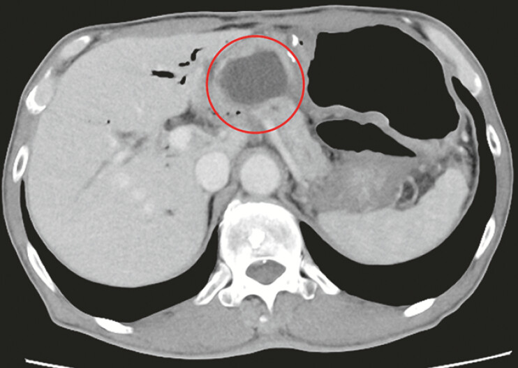 Fig. 1
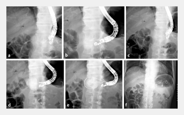 Fig. 2
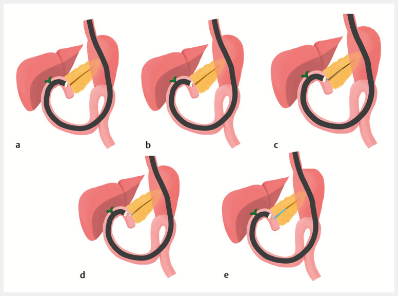 Fig. 3
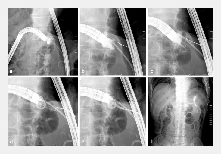 Fig. 4
Peer Reviews
No public reviews on file for this paper yet. If you reviewed it on a platform where reviews are public (OpenReview, ICLR, NeurIPS, ICML), you can paste yours below so the community can read it here.
Videos
No videos yet. Explain this paper in a talk, walkthrough, or lecture? Add one.
Taxonomy
TopicsPancreatic and Hepatic Oncology Research · Gallbladder and Bile Duct Disorders · Gastrointestinal disorders and treatments
Pancreaticojejunal anastomotic strictures (PJAS) and pancreatic fluid leakage can occur after pancreaticoduodenectomy 1 2 . Treatments include endoscopic retrograde pancreatography using a balloon enteroscope or transgastric endoscopic ultrasound (EUS)-guided procedures 2 3 . These treatments can be challenging in patients with severe or complete anastomotic obstruction. Alternatively, EUS-guided pancreaticojejunostomy (EUS-PJS) allows direct access to the pancreatic duct 4 , and a forward-viewing echoendoscope (FV-EUS; TGF-UCT260J; Olympus Medical Systems) expands the field of view, facilitating the precise identification of and access to the anastomotic site 4 . Herein, we describe our experience implementing EUS-PJS.
A 76-year-old man presented with PJAS and pancreatic fluid leakage 8 months after pancreaticoduodenectomy ( Fig. 1 ). Endoscopic treatment was planned, but single-balloon enteroscopy could not identify the pancreaticojejunostomy site. An EUS-guided rendezvous technique ( Fig. 2 a ) was attempted, but neither a guidewire (GW) nor contrast medium could pass through the pancreaticojejunostomy site; both advanced into the pancreatic fluid leakage area ( Fig. 2 b, c ). Transgastric EUS-guided drainage was performed for pancreatic fluid leakage on the same day ( Fig. 2 d ). Subsequently, EUS-PJS was performed using FV-EUS ( Fig. 3 , Video 1 ), which was advanced to the pancreaticojejunostomy site. The pancreatic duct was identified using EUS, punctured with a 19 G needle (EZ shot3; Olympus Medical Systems), and confirmed with contrast medium ( Fig. 4 a ), then a 0.025-inch GW was inserted ( Fig. 4 b ). The double-GW technique was employed due to significant angulation of the pancreatic duct, and dilation was performed using a drill-type dilator ( Fig. 4 c ). A double-lumen catheter was inserted while retaining 0.035 inches of GW in the pancreatic duct ( Fig. 4 d ). A 3-mm balloon dilator was used to dilate the pancreaticojejunostomy site ( Fig. 4 e ). A 7Fr 5-cm plastic stent was placed to complete the procedure ( Fig. 4 f ). Postoperative adverse events did not occur. EUS-PJS can treat PJAS if balloon enteroscopy or a transgastric EUS-guided approach is unsuccessful.
Pancreatic fistula (red circle) near the pancreaticojejunostomy site.
Endoscopic ultrasound-guided rendezvous technique and EUS-guided pancreatic fistula drainage. a Pancreatic duct puncture via the stomach and contrast medium injection. b Only the pancreatic fistula is visualized from the pancreatic duct. c Guidewire advancement stops at the pancreaticojejunostomy site and advances only to the pancreatic fistula. d Puncture of and contrast medium injection into the pancreatic fistula via transgastric using EUS. e Guidewire placement in the pancreatic fistula. f Transgastric stent placement in the pancreatic fistula. Abbreviation: EUS, endoscopic ultrasound.
Schema of EUS-guided pancreaticojejunostomy. a Insertion of a forward-viewing echoendoscope into the pancreaticojejunostomy site. b Pancreatic duct puncture. c Guidewire placement in the pancreatic duct. d Puncture site dilation. e Stent placement in the pancreatic duct. Abbreviation: EUS, endoscopic ultrasound.
Endoscopic ultrasound-guided pancreaticojejunostomy. a Insertion of a forward-viewing echoendoscope into the pancreaticojejunostomy site, followed by pancreatic duct puncture using a 19G needle and contrast medium injection. b Guidewire placement in the pancreatic duct. c Puncture site dilation using a drill-type dilator. d Placement of two guidewires using a double-lumen catheter. e Puncture site dilation with a 3-mm balloon dilator. f Stent placement in the pancreatic duct. Abbreviation: EUS, endoscopic ultrasound.
Endoscopic ultrasound (EUS)-guided pancreaticojejunostomy after unsuccessful attempts with a balloon enteroscope and a transgastric EUS-guided approach.Video 1
Endoscopy_UCTN_Code_TTT_1AS_2AI
The reference list from the paper itself. Each links out to its DOI / PubMed record.
- 1Cioffi JL Mc Duffie LA Roch AM Pancreaticojejunostomy stricture after pancreatoduodenectomy: outcomes after operative revision J Gastrointest Surg 20162029329910.1007/s 11605-015-3012-z 26553264 · doi ↗ · pubmed ↗
- 2Nabeshima T Kanno A Masamune A Successful endoscopic treatment of severe pancreaticojejunostomy strictures by puncturing the anastomotic site with an EUS-guided guidewire Intern Med 20185735736229151507 10.2169/internalmedicine.9133-17PMC 5827316 · doi ↗ · pubmed ↗
- 3Imoto A Ogura T Higuchi K Endoscopic ultrasound-guided pancreatic duct drainage: techniques and literature review of transmural stenting Clin Endosc 20205352553410.5946/ce.2020.17332967409 PMC 7548157 · doi ↗ · pubmed ↗
- 4Sadek A Hara K Okuno N Safety and efficacy of trans-afferent loop endoscopic ultrasound-guided pancreaticojejunostomy for post pancreaticoduodenectomy anastomotic stricture using the forward-viewing echoendoscope: a retrospective study from Japan. Epub ahead of print.Clin Endosc 202410.5946/ce.2024.089PMC 1198313439188116 · doi ↗ · pubmed ↗