Macular thickness and vascular density assessment using optical coherence tomography and optical coherence tomography angiography imaging in iron ore mine personnel
Navid Faraji, Seyyed Pouria Tafti, Niloofar Khoshroo, Alireza Khoshrou, Elham Bakhtiari, Saeid Eslami, Nasser Shoeibi, Mohammad Reza Ansari Astaneh, Seyedeh Maryam Hosseini, Majid Abrishami, Hamid Reza Heidarzadeh, Parnian Arjmand, Mojtaba Abrishami

TL;DR
This study used OCT and OCTA imaging to assess retinal changes in iron ore mine workers but found no significant differences compared to a control group.
Contribution
The study is the first to investigate retinal structural and vascular changes in iron ore mine workers using OCT and OCTA.
Findings
No significant difference in foveal thickness between mine workers and controls.
Vessel density in retinal capillary plexus showed no significant variation between groups.
Occupational exposure in iron ore mines was not associated with retinal structural changes.
Abstract
To assess macular anatomical and vascular parameters in individuals working in iron ore mines using Optical Coherence Tomography (OCT) and Optical Coherence Tomography Angiography (OCTA) imaging to explore potential correlations between this occupational exposure and retinal changes. Individuals from the Sangan iron ore mine in Iran were included in a comparative cross-sectional observational study. An age-matched normal control group was selected from healthy participants employed at Mashhad University of Medical Sciences. Following thorough medical evaluations, participants underwent OCT and OCTA imaging. The macular thickness profile, vessel density (VD) of the superficial (SCP) and deep retinal capillary plexus (DCP), and the area of the foveal avascular zone (FAZ) were measured in our cases and compared with age-matched normal controls. One hundred and one individuals, with an…
Genes, proteins, chemicals, diseases, species, mutations and cell lines named across the full text — each resolved to its canonical identifier and authoritative record.
Click any figure to enlarge with its caption.
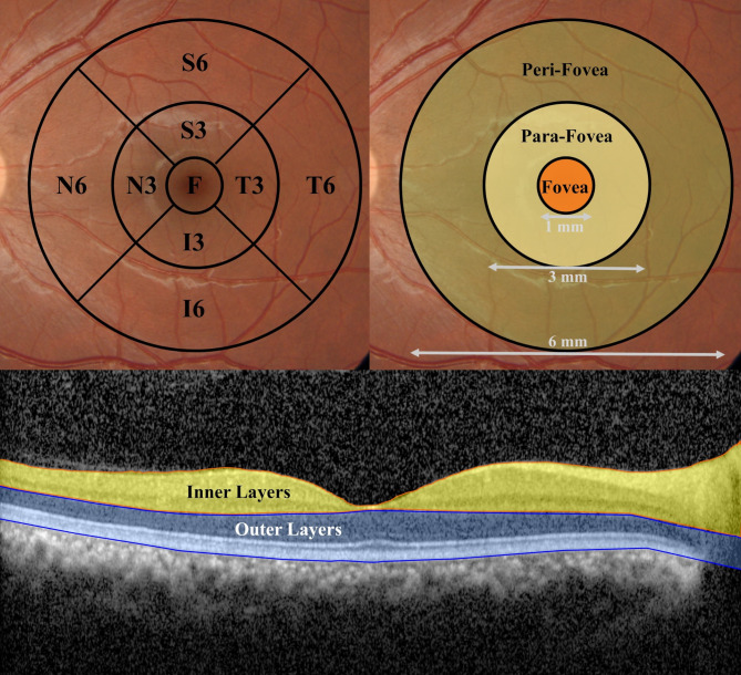 Figure 1
 Figure 2
Peer Reviews
No public reviews on file for this paper yet. If you reviewed it on a platform where reviews are public (OpenReview, ICLR, NeurIPS, ICML), you can paste yours below so the community can read it here.
Videos
No videos yet. Explain this paper in a talk, walkthrough, or lecture? Add one.
Taxonomy
TopicsRetinal Imaging and Analysis · Retinal Diseases and Treatments · Retinal and Optic Conditions