Corrigendum: Metabolic-imaging of human glioblastoma live tumors: a new precision-medicine approach to predict tumor treatment response early
Mariangela Morelli, Francesca Lessi, Serena Barachini, Romano Liotti, Nicola Montemurro, Paolo Perrini, Orazio Santo Santonocito, Carlo Gambacciani, Matija Snuderl, Francesco Pieri, Filippo Aquila, Azzurra Farnesi, Antonio Giuseppe Naccarato, Paolo Viacava, Francesco Cardarelli

Abstract
Genes, proteins, chemicals, diseases, species, mutations and cell lines named across the full text — each resolved to its canonical identifier and authoritative record.
Click any figure to enlarge with its caption.
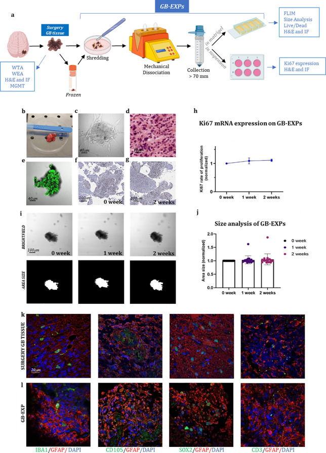 Figure 1
Peer Reviews
No public reviews on file for this paper yet. If you reviewed it on a platform where reviews are public (OpenReview, ICLR, NeurIPS, ICML), you can paste yours below so the community can read it here.
Videos
No videos yet. Explain this paper in a talk, walkthrough, or lecture? Add one.
Taxonomy
TopicsCancer, Hypoxia, and Metabolism · Medical Imaging Techniques and Applications · Metabolomics and Mass Spectrometry Studies
In the published article, there was an error in Figure 1 as published. The two figures above and below in Figures 1K-L, related to SOX2 staining, are part of a series of photographs aimed at demonstrating that GB explants—small tissue fragments approximately 300 µm in size—retain the original cytoarchitecture of the tissue from which they are derived (surgery tissue). The upper figure represents an image of the surgery tissue, while the lower figure shows the corresponding small explant (GB-EXP) derived from it. The SOX2 staining was specifically performed to add further evidence of the preserved representation of tumor components within the GB explant, a conclusion also supported by other images in the series (GFAP; CD105,CD33).
We acknowledge an error of duplication in the figures related to SOX2, as the upper image is, in fact, a photo of the same explant but with a slightly shifted area. We deeply regret this misplacement and are providing the correct image of the tumor stained with SOX2.The corrected Figure 1 and its legend appear below.
The authors apologize for this error and state that this does not change the scientific conclusions of the article in any way. The original article has been updated.