Fiber architecture in the human ventromedial striatum and its relation with the bed nucleus of the stria terminalis
Oliver Krüger, Uwe Klose, Gisela E. Hagberg, Thomas Shiozawa-Bayer, Henry Evrard, Cintia Meszaros, Thomas Ethofer, Klaus Scheffler, Ulrike Ernemann, Benjamin Bender, Stijn Michielse, Stijn Michielse, Stijn Michielse

TL;DR
This study explores the fiber connections in the human brain's ventromedial striatum and its link to the bed nucleus of the stria terminalis using advanced imaging and microscopy.
Contribution
The study confirms and expands on earlier findings by using polarized light microscopy to reveal detailed fiber architecture in the hCN and NAcc.
Findings
PLM revealed distinct fiber populations in the hCN and NAcc, primarily related to the anterior limb of the internal capsule.
Fibers from the BST were sparsely present in the hCN and either terminated there or joined the ALIC.
DTI data showed interindividual variation in fiber density within the hCN, which was confirmed by PLM.
Abstract
The bed nucleus of the stria terminalis (BST) and the ventromedial striatum (consisting of the head of the caudate nucleus (hCN) and the nucleus accumbens (NAcc)) are both part of complex, foremost limbic networks involved in a variety of neuropsychiatric conditions. However, data on functional or structural connections between the BST and hCN in humans are scarce. In an earlier study using both diffusion tensor magnetic resonance imaging (DTI) and conventional histology we found a pathway from the BST to the orbitofrontal cortex apparently passing directly through the hCN. To confirm this finding, we now examined the hCN in human ex-vivo brain tissue using polarized light microscopy (PLM), a method particularly suitable for depicting myelinated nerve fibers. We further examined whether differences in fiber distribution inside the hCN could be depicted using high-resolution DTI data.…
Genes, proteins, chemicals, diseases, species, mutations and cell lines named across the full text — each resolved to its canonical identifier and authoritative record.
Click any figure to enlarge with its caption.
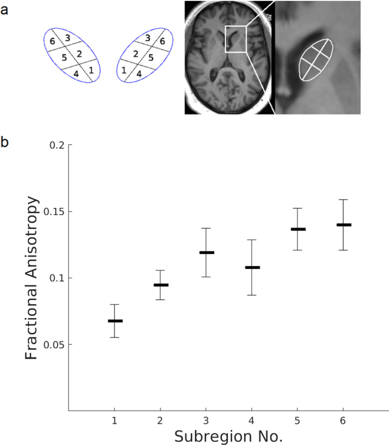 Figure 1
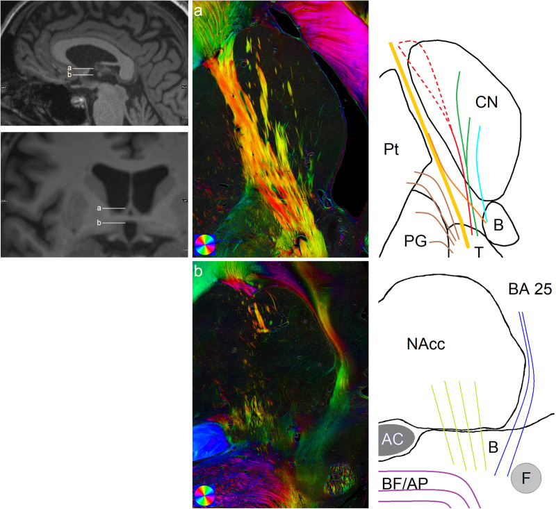 Figure 2
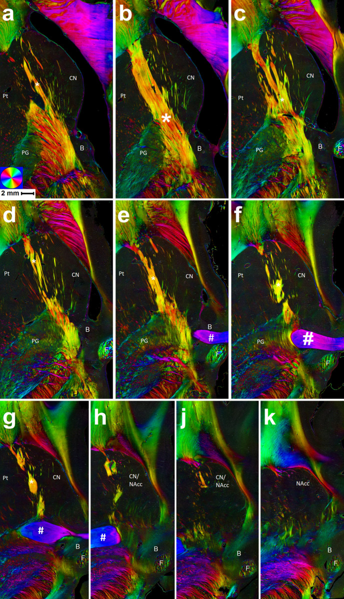 Figure 3
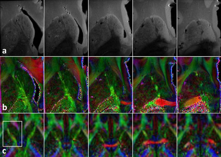 Figure 4
Peer Reviews
No public reviews on file for this paper yet. If you reviewed it on a platform where reviews are public (OpenReview, ICLR, NeurIPS, ICML), you can paste yours below so the community can read it here.
Videos
No videos yet. Explain this paper in a talk, walkthrough, or lecture? Add one.
Taxonomy
TopicsAdvanced Neuroimaging Techniques and Applications · Neurological disorders and treatments · Functional Brain Connectivity Studies