Review on Symptomatic pedunculated leiomyomas in pregnancy with special consideration of an example case
Jonas Bubmann, Carl Mathis Wild, Christian Dannecker, Manuela Franitza, Bernadette Eser, Marina C. Seefried, Thomas Kroencke, Philipp Voisard, Udo Jeschke, Fabian Garrido

TL;DR
This paper reviews cases of large fibroids during pregnancy and shows that surgical removal can be safe and effective.
Contribution
The paper provides a case example and literature review to support myomectomy as a safe option for large pedunculated fibroids in pregnancy.
Findings
Myomectomy is safe for pedunculated fibroids during pregnancy.
Laparotomy is recommended for fibroids larger than 10 cm.
Surgical removal does not preclude vaginal birth in such cases.
Abstract
Symptomatic pedunculated leiomyomas in pregnancy; review of the literature with special consideration of an example case. Retrospective narrative review with an example case. Systematic evaluation of 37 reports. A 36-year-old Caucasian primigravida was referred symptomatic at 16 + 0 weeks due to a 13,5 cm myoma causing pain, constipation, urine retention and dysesthesias. Our patient underwent myomectomy at 17 + 0 weeks. One pedunculated leiomyoma was successfully removed. Myomectomy can be performed and is safe for pedunculated fibroids in pregnancy. Depending on the clinical scenario, surgical removal may be indicated. Based on the size of the fibroids and expected adhesions, a laparotomy is a safe option and is not a contraindication for vaginal birth in the case of pedunculated fibroids. Myomas larger than 10 cm should be removed by laparotomy.
Genes, proteins, chemicals, diseases, species, mutations and cell lines named across the full text — each resolved to its canonical identifier and authoritative record.
Click any figure to enlarge with its caption.
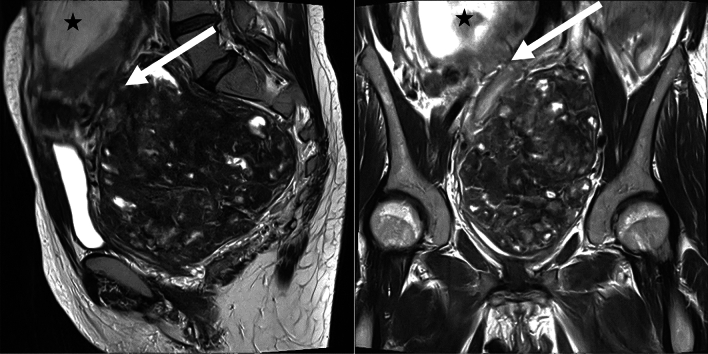 Figure 1
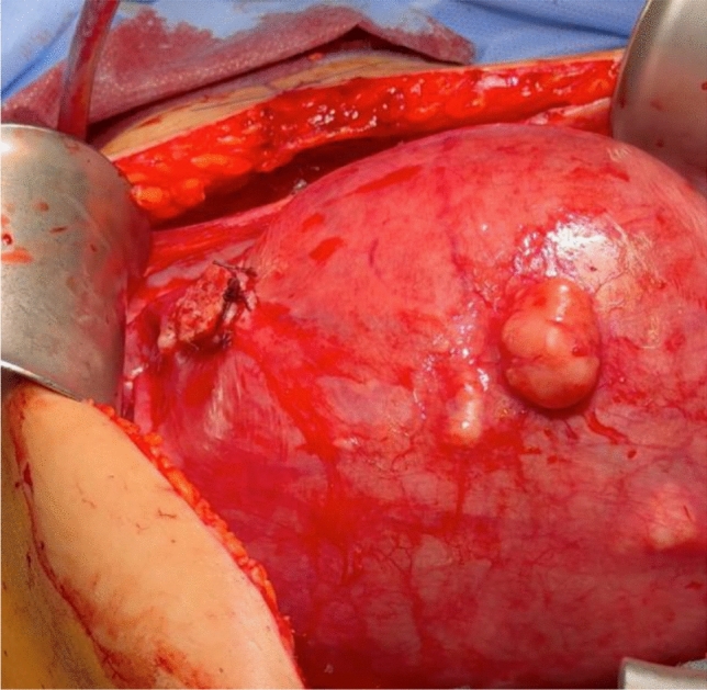 Figure 2
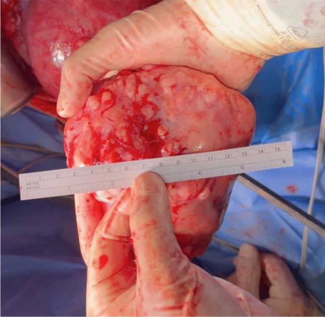 Figure 3
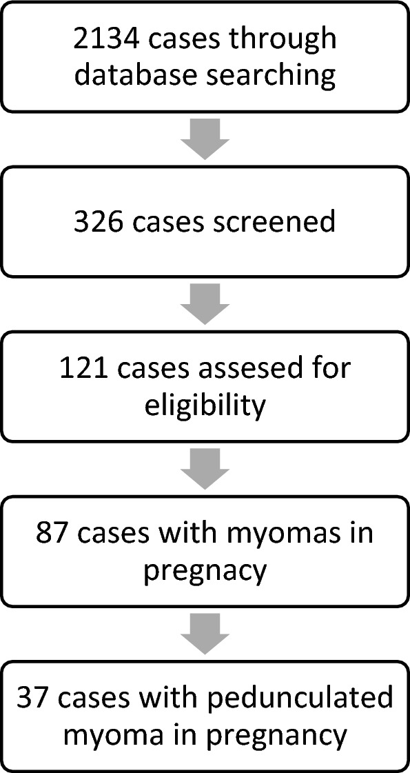 Figure 4
- —Universität Augsburg (3144)
Peer Reviews
No public reviews on file for this paper yet. If you reviewed it on a platform where reviews are public (OpenReview, ICLR, NeurIPS, ICML), you can paste yours below so the community can read it here.
Videos
No videos yet. Explain this paper in a talk, walkthrough, or lecture? Add one.
Taxonomy
TopicsUterine Myomas and Treatments · Minimally Invasive Surgical Techniques · Endometriosis Research and Treatment
Introduction
Leiomyomas are the most common (20–40%) benign disease affecting the female reproductive system. The prevalence of leiomyomas in pregnancy is 2–10% and is usually asymptomatic, but 10% of patients develop complications in pregnancy [1]. Pain is often seen in women with fibroids > 5 cm [2]. In early pregnancy the volume of fibroids increases by 12% in volume because of the rapid increase in serum chorionic gonadotropin levels [3]. Only 37 pedunculated fibroids during pregnancy with single myomectomy are reported in the literature [4].Complication risk does increase with the number of fibroids, size, relation to the placenta and location [4]. Pain is related to the blood supply of the fibroid because increased growth can result in insufficient blood supply followed by necrosis [2].
Example case presentation
A 36-year-old primigravida presented to our University Hospital in January 2024 with a one-year history of chronic hematochezia, which had been treated with oral iron supplementation. The patient had been in the 16 + 0 weeks of gestation with a viable fetus (single, intra-uterine). The patient reported flank pain and micturition disorder. Diagnostic work-up included transabdominal ultrasound which identified a previously unknown pelvic mass. MR imaging was performed for better delineation and characterization of the mass. A 13.5 × 13 cm measuring pedunculated sub-serosal leiomyoma was diagnosed, originating from the posterior wall of the uterus. The lower abdominal organs had been shifted upwards with a compression-related urinary stasis III° on the left side and a compression of the common iliac vein (Fig. 1).Fig. 1. Preoperative MRI. T2-weighted images in the sagittal and coronal plane depicts a pedunculated sub serosal Leiomyoma (arrowhead) and the fetus (asterisk). The vascular pedicle visualized by worm-like signal-free vessels (arrowhead) suggests an attachment side of the leiomyoma to the posterior wall of the uterus. Degenerative changes (white spots) are seen within the leiomyoma
The uterine leiomyoma had not been diagnosed before the pregnancy, because the patient has not undergone regular screenings. The pregnancy had been developing physiological. After interdisciplinary discussion prophylactic low molecular weight heparin was prescribed. The patient developed dysesthesia in the left leg in the following weeks. Because of the increasing symptoms a conservative vs. surgical therapy was discussed with the patient. Double-J catheters had been inserted prior to surgery. Because of declining hemoglobin levels, two erythrocyte concentrates had been transfused.
On 24/01/2024 a longitudinal laparotomy was performed under general anesthesia with endotracheal intubation. Operative findings included normal liver, spleen, kidneys, diaphragm, ovaries and fallopian tubes. The uterus was soft and the size was adequate for 17 + 0 weeks of gestation. Fetal movements were visible. A pedunculated fibroid without a torsion measuring 13,5 cm in diameter filled out the whole lower sacral cavity. The pedicle was originating from the dorsal uterus with a stalk diameter of 2 cm. The fibroid was adherent to the peritoneum and sigmoid colon. It was detached and the pedicle was cut after ligation with several sutures 2-0 vicryl. After myomectomy, the pedicle was found to originate from the anterior wall of the uterus and had been turned during pregnancy dorsally. The estimated blood loss was 400 ml and the time of the surgery was 85 min. The tumor weighted 740 g and was sent for pathology. Pre-, intra- and postoperative performed sonographic vital controls of the fetus revealed normal findings (Fig. 2, 3).Fig. 2. Stump of the pedicleFig. 3Myoma
Postoperatively, the patient was given indomethacin prophylactically to prevent uterine contractions. In the Further course, there was a slight neuropathy of the left leg, which after further diagnostics using MRI was most likely interpreted as the expression of an irritation caused by the pressure of the fibroid. There were no further abnormalities.
The symptoms improved and the patient was discharged from the hospital 10 days after the operation. The histological findings showed a leiomyoma with regressive changes and focal ischemic necrosis. There was no evidence of malignancy. The patient was followed up by us twice, and the pregnancy developed normally with no further complaints. Unfortunately, the patient decided to give birth in a rural hospital at 38 + 1 weeks’ gestation. Due to her history, a spontaneous labor was not performed and a secondary caesarean section was performed. A 3300 g baby with APGAR 9–10-10 was delivered. The patient was discharged after 4 days without complications.
The patient’s written consent to publication has been obtained.
Review of literature
Material and methods
In March 2024, the search was carried out with various search terms in Pubmed^®^ (Table 1). The initial search was for case reports and case descriptions of fibroids during pregnancy. Cases with myomectomy during cesarean section were excluded. The cases were then reduced to pedunculated fibroids. Vaginally pedunculated fibroids were excluded. A table was created and the following parameters were recorded for each case: Year of treatment, name of author, title of publication, age of patient, parity, week of pregnancy at first presentation, week of pregnancy at treatment, location of fibroid, symptoms, sonographic findings, MRI findings, size of fibroid, type of operation, special features of operation, blood loss, antibiotics, tocolysis, pneumoperitoneum, intraoperative torsion, pathological findings, weight of fibroid, time of discharge, type of birth, week of pregnancy at birth and other complications. The data collection was carried out by one author (B.J.) (Fig. 4).Table 1. Presentation of cases used for this reviewAuthorDateAgePregnancyGestational week of surgeryLocation of myomaSymptomsImagingType of operationMyoma weightSize of myomaType of birthWeek of birthMakar et al.198914Posterior wallPelvic painSonography120 mmVaginalKalantaridou et al.19943819FundusPelvic painSonographyLongitudinal laparotomy1500 g37Pelosi et al.199535Primipara13FundusPelvic painSonographyLSK, free morcellation into abdominal cavity1500 g60 mmCesarean section for breech presentation39Sciannameo et al.19963120MRIMajid et al.199735Multipara17FundusGastrointestinal symptomsSonography240 mmIntrauterine fetal death19Luxman et al.199727Primipara15FundusSonographyLSK, free morcellation into abdominal cavity80 mmVaginal39Kalantaridou et al.19992516FundusPelvic painSonography170 g39Wittich et al.200031Primipara15FundusPelvic painSonography + MRILongitudinal laparotomy2074 g205 mmElective cesarean section37Kalantaridou et al.20012516Anterior wallPelvic painSonography + MRI625 g39Sentilhes et al.200335Multipara17Left lateral wallPelvic painSonographyLSK, free morcellation into abdominal cavity50 mmElective cesarean section37Melgrati et al.200529Primipara24FundusPelvic pain, feverSonographyIsobaric Laparoscopy70 mmVaginal39Dracea et al.200639Multipara14FundusSonographyTransverse Laparotomy240 mmVaginal37Usifo et al.200731Primipara13Posterior wallPelvic pain, gastrointestinal symptomsSonographyTransverse Laparotomy168 mmElective cesarean section38Okokwo et al.20074019FundusLower extremity edemasSonographyLongitudinal laparotomy10,000 g280 mmElective cesarean section38Leite et al.200743Primipara17FundusPelvic painSonographyLongitudinal laparotomy91 mmVaginal39Alanis et al.200822Multipara13FundusPelvic painMRILongitudinal laparotomy8000 g300 mmVaginal38Suwandinata et al.200828Primipara15Posterior wallPelvic painSonographyLongitudinal laparotomy320 g80 mmElective cesarean section37Camacho et al.200935Multipara16Posterior wallPelvic pain, feverSonographyLongitudinal laparotomy62 mmVaginal40Bhatla et al.200930Primipara20FundusPelvic pain, gastrointestinal symptomsSonographyLongitudinal laparotomy3900 g280 mmVaginal38Fanfani et al.201039Primipara25FundusPelvic painSonographyLSK, Endo bag extraction95 g90 mmVaginal40Son et al.201131Primipara18Posterior wallSonography + MRILSK, Endo bag extraction108 g90 mmVaginal39Ardovino et al.201131Multipara14FundusPelvic painSonographyLSK, free morcellation into abdominal cavity127 g63 mmVaginal40Pelissier-Komorek et al.201234Primipara10FundusPelvic pain, dyspneaSonography + MRILongitudinal laparotomy2040 g220 mmVaginal35Doerga-Bachasing et al.201233Multipara10Posterior wallPelvic pain, gastrointestinal symptomsSonography + MRILongitudinal laparotomy175 mmCesarean section40Macció et al.201233Primipara19FundusPelvic pain, gastrointestinal symptoms, vaginal bleedingSonographyLSK, Endo bag extraction250 g150 mmElective cesarean section with FGR39Macció et al.201224Primipara20FundusPelvic painSonographyLSK, Endo bag extraction170 g100 mmVaginal40Macció et al.201234Primipara20Anterior wallPelvic painSonographyLSK, Endo bag extraction240 g40 mmVaginal39Tabandeh et al.201230Primipara24FundusPelvic pain, gastrointestinal symptomsSonography + MRILongitudinal laparotomy230 mmElective cesarean section37Currie et al.201327Primipara11Anterior wallPelvic painSonographyLSK with Pfannenstiel incision80 mmDomenici et al.201335Primipara16Posterior wallPelvic pain, urinary habit changesSonography + MRILongitudinal laparotomy200 mmElective cesareans section38Saccardi et al.201435Primipara15Anterior wallPelvic pain, gastrointestinal symptomsSonographyLSK, free morcellation into abdominal cavity1363 g240 mmCesarean section for fetal tachycardia41Anthimides et al.201531Multipara10FundusPelvic painSonographyLSK, Endo bag extraction77 mmAlgara et al.20153618Pelvic painLSKIntrauterine fetal death after car accidentJhalta et al.201634Primipara14FundusSonographyLongitudinal laparotomy160 mmVaginal39Kim et al.201635Primipara10FundusPelvic painSonographyLSK93 mmVaginal41Basso et al.201736Multipara17Anterior wallPelvic painSonographyLongitudinal laparotomy132 mmVaginal38Our case202437Primipara18Anterior wallPelvic pain, gastrointestinal symptoms, urinary habit changesSonography + MRILongitudinal laparotomy740 g135 mmSecondary caesarean section39Fig. 4Overview of the review process
Statistics
The statistical analysis was performed using data from eligible studies to assess the overall effect sizes and heterogeneity. Effect measures, such as odds ratios (OR) and risk ratios (RR), with 95% confidence intervals (CI), were calculated for binary outcomes, while mean differences (MD) were used for continuous outcomes. Funnel plots and Egger’s regression test were conducted to assess potential publication bias, with a p-value < 0.05 considered statistically significant.
Results
2134 cases were found and 326 of them further screened. 121 cases had been eligible, but only 87 of them had myomectomies during pregnancy. All cases before the introduction of ultrasound diagnostics were excluded. The oldest included case was from 1989 [5], seven cases were before 2000 [6–10], twelve cases between 2000 and 2010 [6, 11–21] and 17 cases since 2010 to date [22–36]. The most recent case was from 2017 [36]. 37 cases reported about pedunculated myomas during pregnancy. One study was translated into Spanish. The remaining cases were in English. We include our above-mentioned case in the systematic review.
Demography
The age was noted in 37 [6–23, 23–36] of the 38 cases. The average age was 32.7 years (± 4.72; 22–43 years). 55.3% (n = 21) were primipara [7, 10, 11, 13, 15, 17, 19, 21–23, 26, 28–31, 34, 35] and 26,3% (n = 10) of the patients had already been pregnant once [9, 12, 14, 18, 20, 25, 27, 32, 36], whereby no information was provided in 7 case reports [5, 6, 8, 16, 33].
Symptomatology and diagnosis
On average, the patients presented at 14.97 weeks of pregnancy (± 4.38; range: 7–25). Only one patient [14] showed no symptoms at all and no information about the symptoms was reported in three patients [8, 10, 24]. 81.6% (n = 31) of the patients reported pelvic pain. In addition, 18.4% (n = 7) had gastrointestinal symptoms in combination or alone [9, 15, 21, 22, 27–29]. Urinary habit changes were reported in 5.3% (n = 2) of cases [31]. Fever, edema of the lower extremities or vaginal bleeding were also described in 2.6% (n = 1) [16, 20, 28].
71.1% (n = 27) of the patients had only a preoperative sonography [5–7, 9, 10, 12–17, 19–23, 25, 28, 30, 32, 34–36], with 5,3% (n = 2) having an MRI [8, 18] and only 18,4% (n = 7) having an MRI and sonography [11, 26–29, 31]. No examination was documented in one patient [33].
A pedunculated fibroid was diagnosed preoperatively in 14 ultrasound findings [5, 6, 14, 22, 25, 27, 28, 35], with 22 findings not describing a pedicle. 55.3% (n = 21) of the fibroids originated from the fundus, [6, 7, 9–11, 13, 14, 16–18, 21, 23, 25, 26, 28, 29, 32, 34, 35] followed by 21.1% (n = 8) from the posterior wall [5, 15, 19, 20, 27, 28, 31]. 15.8% (n = 6) were on the anterior wall [22, 28, 30, 36] and only one fibroid (2.6%) was lateral to the uterus [12].
A sub-serosal fibroid was described in all MRI examinations, [8, 11, 17, 24, 27, 29, 31] whereby a torsion could already be detected in one finding [8]. At least one further fibroid was diagnosed in 28,9% (n = 11) of the patients [6, 9, 14, 19, 26, 28, 31, 36]. 21% (n = 8) of patients showed multiple uterine fibroids [6, 9, 26, 28, 36].
Management
No case report could be found that describes a wait-and-see approach.
47.4% (n = 18) of the patients underwent open surgery, [6, 11, 14–21, 26, 27, 29, 31, 34, 36] with a longitudinal laparotomy being performed in 42.1% (n = 16) of cases [6, 11, 16–21, 26, 27, 29, 31, 34, 36] and a transverse laparotomy in 5.3% (n = 2) [14, 15]. Laparoscopy (LSK) was performed in 39.5% (n = 15) [7, 10, 12, 13, 22–25, 28, 30, 32, 33, 35].
Significantly (p = < 0.001), larger fibroids underwent laparotomy and smaller fibroids underwent LSK. There was also a significance (p = 0.018) between the weight of the fibroid and the type of surgery. Patients who underwent open surgery had fibroids that were on average 181.71 mm (± 71.64 mm; 62–300 mm) in size and 3571.8 g (± 3556 g; 320–10.000 g) in weight. In contrast, the patients with LSK had 91.6 cm (± 50.13 cm; 40–240) measuring fibroids and lighter fibroids weighing 481.6 g (± 590.1 g; 95–1500 g). Surprisingly, the transverse laparotomy was more likely to yield heavy fibroids (204 g; ± 50.9 g) than longitudinal laparotomy (178.8 g; ± 78.4 g).
In only 8 patients the time between the first presentation and the surgical treatment documented [11, 18, 20–22, 27, 29]. For these, the average time span was 6.25 weeks (± 4.13; 1–12 weeks). We assume that the weeks of pregnancy stated in the case report also correspond to the time of surgical treatment. Thus, on average, the patients underwent surgical intervention at 16.3 weeks of pregnancy (± 3.78; 10–25). Blood loss was reported in 17 patients, [7, 9, 13, 15, 16, 19, 21, 22, 25, 27, 28, 28, 31, 35] with a mean value of 607 ml (± 1.076 ml; 0–4.500 ml) and thus a very wide range. Nevertheless, only one patient is described as requiring a postoperative transfusion [21]. No antibiotics were discussed in 17 patients, [5, 6, 8, 10, 13, 15–17, 26–29, 33] so that 26.3% (n = 10) received antibiotics [12, 14, 18, 19, 21, 31, 34, 36] and 28.9% (n = 11) did not receive antibiotics [8, 10, 12, 21, 23–25, 30, 32]. Only one patient was described as having a post-operative infection with abscess development at the uterine scar. [12] This one patient had a laparoscopy and did not receive antibiotics intraoperatively [12].
26.3% (n = 10) patients received tocolysis pre- or postoperatively, [9, 11, 14, 17, 21, 29, 34, 36]. whereas 34.2% (n = 13) did not receive tocolysis [7, 12, 18–20, 22–25, 30–32, 35]. In 15 patients no information regarding tocolysis was documented [5, 6, 8, 10, 13, 15, 16, 26–28, 33]. No patient developed labor until postoperative discharge. Torsion of the fibroid was present in 26.3% (n = 10) of the cases. [9, 11, 15, 21, 30, 32, 33, 35], However, the majority of 55.3% (n = 21) had no torsion [6, 7, 11–13, 17, 19, 21–23, 25, 27–29, 31, 34]. Of the LSK patients, 4 underwent free morcellation into the abdominal cavity [7, 10, 12, 22, 25]. In 6 of the LSK patients, the fibroid was retrieved using an Endo bag [23, 24, 28, 32]. In one patient, attention was paid to isobaric pressure [13]. The intra-abdominal pressure during LSK was described in 11 patients [7, 10, 13, 19, 22–25, 28, 32] and was found to be 11.2 mmHg (± 1.687 mmHg; 10–14 mmHg) on average.
Histopathology
In 31 operations, a histopathological examination was subsequently performed [5, 7, 9, 11–13, 15–26, 28, 30–36]. This revealed a degenerative change in the fibroid in 41.9% (n = 13) of the patients [6, 12–14, 16–20, 26, 28, 35]. 20 fibroids were described as sub serosal [9, 13, 15, 17, 19, 21, 23, 25–28, 30–36], whereby the other cases did not document any such description regarding the localization.
Postoperative time
On average, patients were discharged 4.64 (± 2.99; 1–14) days after surgery.
81.3% (n = 31) had a complication-free postoperative course. Postoperative complications were described in only 10.5% (n = 4) of the patients [12, 21, 27, 36]. One patient developed an abdominal abscess [12], as mentioned above and one patient required a transfusion of two red blood cell coagulates [21]. One patient developed cervical insufficiency in the 21st week of pregnancy [36].
One child died in the immediate post-operative period after multiple myomas had been removed and an appendectomy was performed [9].
Delivery
On average, all other cases had a birth in the 38.6 (± 1.36; 35–41) week of pregnancy. Of these, 47.2% (n = 17) had a vaginal birth [5, 10, 13, 14, 17, 18, 20, 21, 23–26, 28, 34–36]. A cesarean section was performed in 30.6% (n = 11) of cases [7, 11, 12, 15, 16, 19, 22, 27–29, 31]. No case reported a reduced APGAR or postpartum abnormalities. Significantly (p = 0.002), the patients with laparotomy gave birth at 37.9 (± 1.26; 35–40) weeks gestation, whereas the patients with LSK gave birth at 39.4 (± 1.08; 37–41) weeks gestation. There was no significance in regard to the type of birth.
Discussion
Surgical interventions are avoided during pregnancy, if possible, but our case and review of the literature show that surgical interventions may be necessary in pregnant patients. Symptomatic pedunculated fibroids rarely occur in pregnancy but require a well prepared and considered treatment. Patients with the combination of sonography and MRI showed the lowest complication rate. This is also confirmed by the fact that visualization of a pedicle enabling a diagnosis of a pedunculated leiomyoma was mostly successful on MRI.
Our decision to perform open surgery was in line with the current literature. We conclude that fibroids larger than 10 cm should be treated by laparotomy. In addition, adhesions with the fibroid should always be considered and expected.
The complexity of our case, as well as the literature review, show that surgically experienced personnel are essential for these procedures. Even if there is a temptation to enucleate further fibroids, the cases to date confirm that only the symptomatic fibroid should be removed. Experienced anesthetists should be present, as blood loss of up to 4.5 L has been described. Patients receiving antibiotic therapy showed no further infection, which was also confirmed in our case. Transfusion is not often needed prior surgery [21]. Ligation of the stalk by means of vicryl sutures is the most commonly applied technique, followed by staplers and bipolar electrosurgical devices. Vaginal delivery mode is seen in single myomectomies by 30–45%, even though there is missing data about the pedunculated situation [37].
It is astonishing that neither in patients with tocolysis nor without tocolysis a labor induction kit was described in any case.
Torsion of a fibroid is rare but should be considered first, as further complications such as necrosis or septicemia can develop.
A primary cesarean section was not recommended in any of the cases. No complications were described in any of the vaginal births. This confirms previous literature that vaginal birth is possible after myomectomy without opening the uterine cavity [38].
Conclusion
Myomectomy can be performed safely for pedunculated fibroids in pregnancy. MRI is helpful for fibroid mapping and maybe considered when sonography is insufficient. Based on the size of the fibroids and expected adhesions, a laparotomy is a safe option and is not a contraindication for vaginal birth in case of pedunculated fibroids. Myomas larger than 10 cm should be removed by laparotomy.
The reference list from the paper itself. Each links out to its DOI / PubMed record.