A rare mushroom-shaped lesion growing in the transverse colon
XiaoBo Liu, Jun Pan, TieYan Wang, ShengBao Li, Yuan-Jun Gao

Abstract
Genes, proteins, chemicals, diseases, species, mutations and cell lines named across the full text — each resolved to its canonical identifier and authoritative record.
Click any figure to enlarge with its caption.
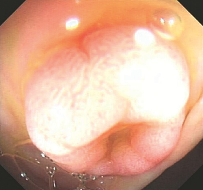 Fig. 1
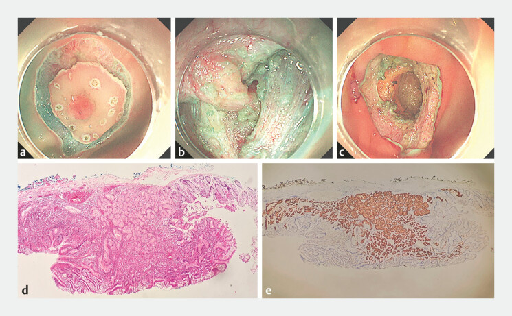 Fig. 2
- —The Health Commission of Hubei Province scientific research project
Peer Reviews
No public reviews on file for this paper yet. If you reviewed it on a platform where reviews are public (OpenReview, ICLR, NeurIPS, ICML), you can paste yours below so the community can read it here.
Videos
No videos yet. Explain this paper in a talk, walkthrough, or lecture? Add one.
Taxonomy
TopicsBiliary and Gastrointestinal Fistulas · Gastrointestinal disorders and treatments · Soft tissue tumor case studies
A 48-year-old woman visited our outpatient department after an outpatient colonoscopy revealed a mushroom-shaped protrusion of Yamada III type in the transverse colon, approximately 60 cm from the anus. It had a diameter of about 8 mm and no surface erosion. However, the center had a shallow depression ( Fig. 1 ). Her outpatient pathological report suggested that the tissue collected was heterotopic gastric mucosa (HGM). When we used an injection needle for submucosal injection, the surrounding area of the tumor was easily lifted, but lifting of the central depression was unsatisfactory ( Fig. 2 a ). After peeling off the mucosal and submucosal layers, we found that this lesion had a thick pedicle, and the root of this pedicle was located in the intrinsic muscle layer and grew toward the serosal layer ( Fig. 2 b , Video 1 ). The postoperative sample looked like a complete mushroom, with a size of about 2.0 × 2.5 cm. After completely removing the lesion, a droplet-shaped pit was left in its original position ( Fig. 2 c ).
Endoscopic appearance of the colonic lesion: a mushroom-shaped protrusion.
Endoscopic submucosal dissection (ESD) and postoperative pathological results. a The surrounding area of the tumor was easily lifted, but lifting of the central depression was unsatisfactory. b The root of the pedicle was located in the intrinsic muscle layer and grew toward the serosal layer. c A droplet-shaped pit was left in its original position. d Heterotopic gastric mucosa in the ESD postoperative specimen (hematoxylin and eosin, original magnification ×10). e Immunohistochemistry confirmed positivity for MUC6.
Endoscopic submucosal dissection for removal of gastric mucosal ectopia originating from the intrinsic muscle layer of the transverse colon.Video 1
Under hematoxylin and eosin staining microscopy, ectopic gastric mucosal tissue was observed on the left and middle parts, presenting as a polyp-like protrusion with a surface covered by gastric pit epithelium. The right side had a colonic mucosal epithelium rich in goblet cells, and the crypt structure was preserved ( Fig. 2 d ). Immunohistochemistry showed that MUC5AC was expressed in the gastric pit epithelium and MUC2 was expressed in the normal colonic mucosal epithelium. MUC6 was expressed in the gastric pyloric glandular tissue ( Fig. 2 e ), confirming HGM in the transverse colon.
HGM can appear throughout the digestive tract and is relatively rare in the colon. Val-Bernal et al. 1 reported a case of a polyp located 50 cm from the anal margin, and Ito et al. 2 reported a case of colonic ectopic gland, a submucosal tumor located in the transverse colon with a raised center surrounded by a fissure. To the best of our knowledge, our study is the first case of HGM with a mushroom-like appearance appearing in the transverse colon, and the mass affected almost the entire intestinal wall.
Endoscopy_UCTN_Code_TTT_1AQ_2AD_3AD
The reference list from the paper itself. Each links out to its DOI / PubMed record.
- 1Val-Bernal J Cagigal M Mayorga M Colonic tubular adenoma with incidental oxyntic gastric heterotopia Rom J Morphol Embryol 20216231331834609438 10.47162/RJME.62.1.35PMC 8597387 · doi ↗ · pubmed ↗
- 2Ito S Hotta K Imai K Endoscopic submucosal dissection for a heterotopic gland in the transverse colon Endoscopy 201749 E 288E 28910.1055/s-0043-11794228905334 · doi ↗ · pubmed ↗