Dual-frequency intraductal ultrasonography: a breakthrough in biliopancreatic imaging during endoscopic retrograde cholangiopancreatography
Yao Lu, Xiaoyan Lv, Shun He

Abstract
Genes, proteins, chemicals, diseases, species, mutations and cell lines named across the full text — each resolved to its canonical identifier and authoritative record.
Click any figure to enlarge with its caption.
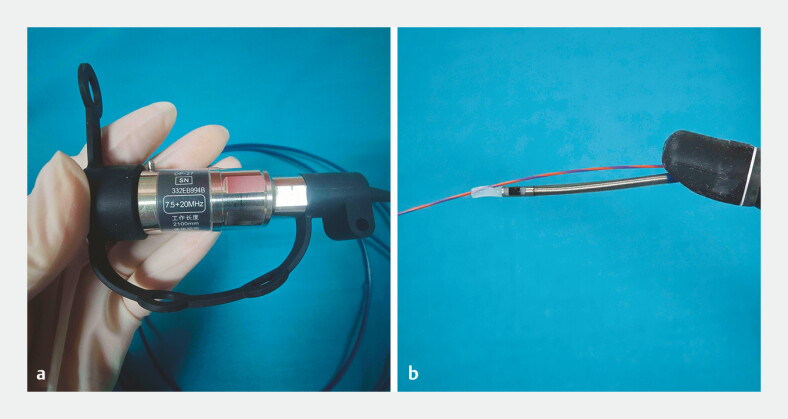 Fig. 1
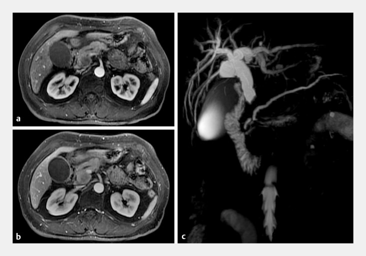 Fig. 2
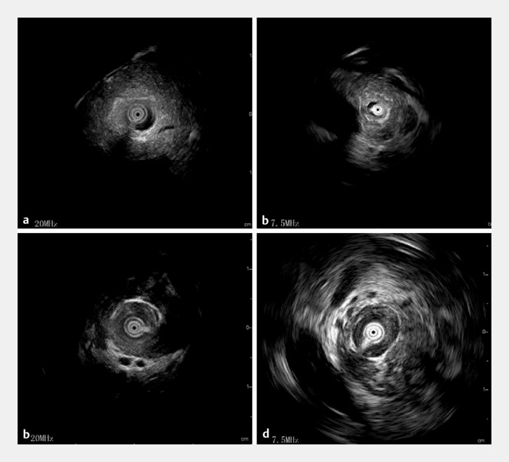 Fig. 3
Peer Reviews
No public reviews on file for this paper yet. If you reviewed it on a platform where reviews are public (OpenReview, ICLR, NeurIPS, ICML), you can paste yours below so the community can read it here.
Videos
No videos yet. Explain this paper in a talk, walkthrough, or lecture? Add one.
Taxonomy
TopicsGallbladder and Bile Duct Disorders · Pancreatic and Hepatic Oncology Research · Cholangiocarcinoma and Gallbladder Cancer Studies
Intraductal ultrasonography (IDUS) is a reliable procedure for evaluating the biliopancreatic duct during endoscopic retrograde cholangiopancreatography (ERCP) 1 2 ; however, conventional high frequency IDUS is limited by its penetration depth 3 . This case highlights a novel dual-frequency IDUS probe that overcomes this limitation ( Fig. 1 ; Video 1 ).
Photograph of the dual-frequency intraductal ultrasonography probe showing: a the probe, which features two frequencies (20 MHz and 7.5 MHz) that are switchable via the main engine; b the probe tip, which has an outer diameter of 2.5 mm.
A novel dual-frequency intraductal ultrasonography probe is used to evaluate biliopancreatic disease during endoscopic retrograde cholangiopancreatography.Video 1
A 60-year-old woman was referred to our hospital with jaundice. Magnetic resonance imaging (MRI) revealed a pancreatic head mass with distal bile duct obstruction ( Fig. 2 ). Laboratory tests showed she had a serum total bilirubin of 330 μmol/L and a CA19-9 of 146 U/mL, and a preliminary clinical diagnosis of pancreatic head cancer was made.
Enhanced magnetic resonance imaging and magnetic resonance cholangiopancreatography (MRCP) images showing: a in arterial phase, mild enhancement of the pancreatic head lesion; b in portal phase, progressive enhancement; c on MRCP, a distal bile duct stricture with upstream bile duct dilatation and slight pancreatic duct dilatation.
Endoscopic retrograde cholangiography (ERC) revealed a defect in the distal bile duct on contrast injection. To determine the nature of this biliary stricture, a novel IDUS probe with dual frequencies of 20 MHz and 7.5 MHz (DP-27, 7.5+20 MHz; Innermed, Shenzhen, China) was advanced over guidewires into the pancreatic duct and bile duct, which were scanned using the pull-back method. Using the 20-MHz frequency, the IDUS scan showed the pancreatic duct and proximal surrounding structures ( Fig. 3 a ). On switching to 7.5 MHz, the far-field resolution significantly improved, allowing visualization of the complete pancreatic contour and parenchyma ( Fig. 3 b ). The pancreatic head appeared as a heterogeneous hypoechoic region without any evident tumorous lesions. A subsequent 20-MHz scan of the intrapancreatic bile duct revealed a circular symmetrical wall thickening, with a smooth outer margin ( Fig. 3 c ). A switch to 7.5 MHz confirmed no evidence of an extrinsic lesion causing compression ( Fig. 3 d ).
Images during dual-frequency intraductal ultrasonography (IDUS) showing; a the appearance on 20-MHz IDUS of the pancreatic duct; b on 7.5-MHz IDUS, a high resolution image of the pancreatic parenchyma; c on 20-MHz IDUS, intrapancreatic biliary wall thickening; d on 7.5-MHz IDUS, detailed periductal structural information.
The IDUS images were therefore used to make a diagnosis of autoimmune pancreatitis and IgG4-related sclerosing cholangitis (IgG4-SC), which was confirmed by finding an elevated serum IgG4 level and on endoscopic ultrasound-guided fine-needle aspiration (EUS-FNA). Following glucocorticoid therapy, the patient’s symptoms resolved, and imaging showed significant improvement. This dual-frequency IDUS technology offers enhanced diagnostic capability and can be seamlessly integrated into routine ERCP procedures, significantly reducing diagnostic delays and improving patient management.
Endoscopy_UCTN_Code_TTT_1AS_2AD
The reference list from the paper itself. Each links out to its DOI / PubMed record.
- 1Chen L Lu Y Wu JC Diagnostic utility of endoscopic retrograde cholangiography/intraductal ultrasound (ERC/IDUS) in distinguishing malignant from benign bile duct obstruction Dig Dis Sci 20166161061726423079 10.1007/s 10620-015-3896-1 · doi ↗ · pubmed ↗
- 2Naitoh I Nakazawa T Endoscopic retrograde cholangiopancreatography and intraductal ultrasonography in the diagnosis of autoimmune pancreatitis and Ig G 4-related sclerosing cholangitis J Med Ultrason 20214857358010.1007/s 10396-021-01114-134331625 · doi ↗ · pubmed ↗
- 3Cheon YK Intraductal ultrasonography for biliary strictures Clin Endosc 20235616416810.5946/ce.2022.18436800766 PMC 10073858 · doi ↗ · pubmed ↗