Exposure of an obstructed splenic artery aneurysm stent into the duodenum
Yutaka Ogikubo, Shun Osumi, Shosuke Hosaka, Kazushi Fukagawa, Satoshi Ono

Abstract
Genes, proteins, chemicals, diseases, species, mutations and cell lines named across the full text — each resolved to its canonical identifier and authoritative record.
Click any figure to enlarge with its caption.
 Fig. 1
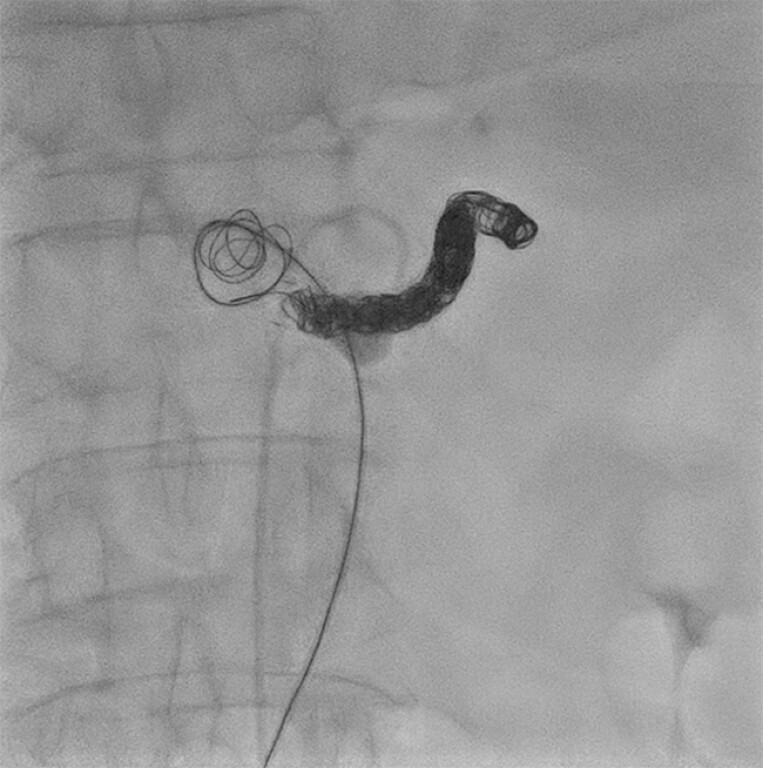 Fig. 2
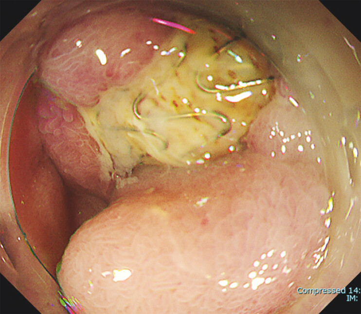 Fig. 3
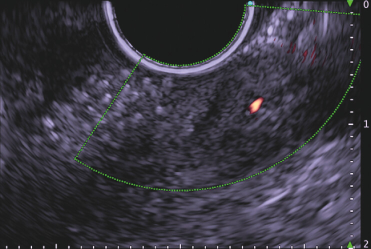 Fig. 4
Peer Reviews
No public reviews on file for this paper yet. If you reviewed it on a platform where reviews are public (OpenReview, ICLR, NeurIPS, ICML), you can paste yours below so the community can read it here.
Videos
No videos yet. Explain this paper in a talk, walkthrough, or lecture? Add one.
Taxonomy
TopicsAbdominal vascular conditions and treatments · Esophageal and GI Pathology · Vascular anomalies and interventions
A 76-year-old man with a history of splenic artery aneurysm and celiac artery aneurysm was referred for a screening esophagogastroduodenoscopy (EGD). An endovascular stent had been placed for the splenic artery aneurysm 5 years previously ( Fig. 1 ), and coiling had been performed in the stent lumen 4 months previously 1 ( Fig. 2 ).
Computed tomography image showing an endovascular stent that had been placed in a splenic artery aneurysm.
Fluoroscopic image showing a coil within the stent lumen.
EGD revealed exposure of a metal mesh in the duodenum, which was thought to be the endovascular stent that had been placed in a splenic artery aneurysm ( Fig. 3 ); no obvious bleeding was observed. Contrast-enhanced computed tomography was performed, but blood flow within the stent could not be evaluated owing to artifacts caused by the stent, and detailed observation was also difficult with transabdominal ultrasonography 2 . Therefore, endoscopic ultrasonography (EUS) was performed from within the stomach, taking care not to directly affect the exposed stent, and we were able to evaluate the stent through the metal mesh, with acoustic shadow but no obvious blood flow seen in the lumen on Doppler echo 3 ( Fig. 4 ; Video 1 ).
Endoscopic image showing a metal mesh exposed in the duodenum.
Endoscopic ultrasonography image showing no obvious blood flow in the stent lumen.
Esophagogastroduodenoscopy shows an exposed metal mesh in the duodenum, which was thought to be an endovascular stent placed in a splenic artery aneurysm; the stent was evaluated with endoscopic ultrasonography through the metal mesh, with acoustic shadow but no evidence of luminal blood flow on Doppler echo.Video 1
Surgical removal of the exposed stent was considered, but the patient was judged to be unsuitable for this owing to his severe co-morbidities. Instead, observation was chosen because of the fact that he had been able to manage oral intake without any problems.
Endoscopy_UCTN_Code_CCL_1AB_2AZ_3AZ
The reference list from the paper itself. Each links out to its DOI / PubMed record.
- 1Abbas MA Stone WM Fowl RJ Splenic artery aneurysms: two decades experience at Mayo clinic Ann Vasc Surg 20021644244910.1007/s 10016-001-0207-412089631 · doi ↗ · pubmed ↗
- 2Boschmann H Zimmermann HB Wiechmann T Ruptured splenic artery aneurysm – a rare cause of recurrent gastrointestinal hemorrhages [in German]Med Klin (Munich)20019635135411450587 10.1007/pl 00002215 · doi ↗ · pubmed ↗
- 3Tannoury J Honein K Abboud B Splenic artery aneurysm presenting as a submucosal gastric lesion: A case report World J Gastrointest Endosc 2016849650010.4253/wjge.v 8.i 14.49627499832 PMC 4959943 · doi ↗ · pubmed ↗