Confirmation of endoscopic injection sclerotherapy by mucosal whitening in the treatment of hemorrhage of refractory recurrent tiny esophageal varices
Kazunori Nagashima, Shintaro Yamaguchi, Ryuichi Maki, Fumihiko Urushibara, Tsunehiro Suzuki, Toshimitu Murohisa, Atsushi Irisawa

Abstract
Genes, proteins, chemicals, diseases, species, mutations and cell lines named across the full text — each resolved to its canonical identifier and authoritative record.
Click any figure to enlarge with its caption.
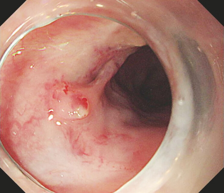 Fig. 1
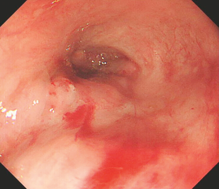 Fig. 2
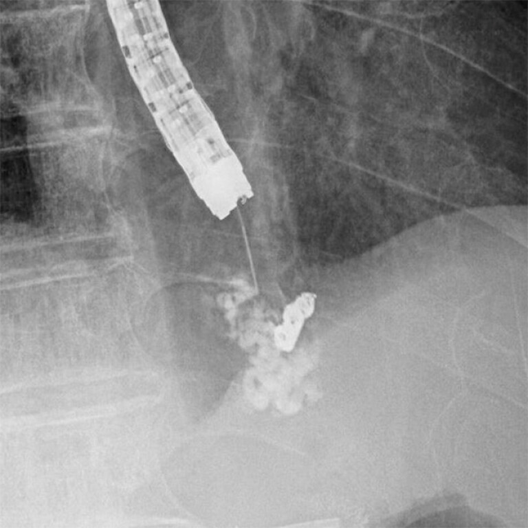 Fig. 3
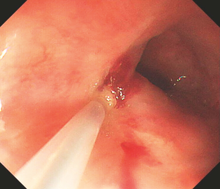 Fig. 4
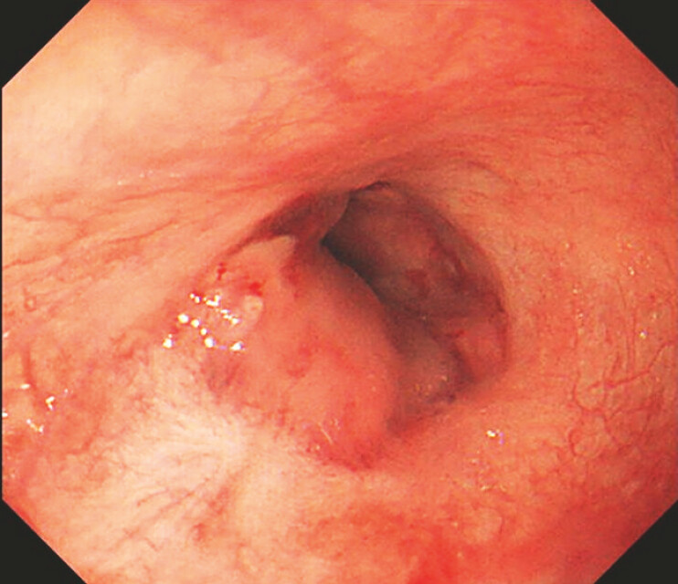 Fig. 5
Peer Reviews
No public reviews on file for this paper yet. If you reviewed it on a platform where reviews are public (OpenReview, ICLR, NeurIPS, ICML), you can paste yours below so the community can read it here.
Videos
No videos yet. Explain this paper in a talk, walkthrough, or lecture? Add one.
Taxonomy
TopicsLiver Disease and Transplantation · Systemic Sclerosis and Related Diseases · Gastrointestinal Bleeding Diagnosis and Treatment
Endoscopic injection sclerotherapy (EIS) is particularly useful for refractory recurrent scarring small varices with red color sign for which endoscopic variceal ligation (EVL) is not feasible. In such cases, intravariceal injection can be confirmed by observing whitening of the mucosa (because of local ischemia) after injection of the sclerosing agent. This report is the first video case confirming that the mucosa around tiny varices whitens during intravariceal EIS.
This video presents a typical case ( Video 1 ). A 75-year-old man with portal vein thrombosis visited our hospital, complaining of hematemesis. He had undergone EVL for esophageal varices in the past. Emergency endoscopy revealed hemorrhagic small esophageal varices with multiple scars caused by EVL ( Fig. 1 , Fig. 2 ). After inflating a balloon attached to the endoscope tip, the varices were punctured using a 25-gauge needle (Varixer; TOP Corp., Tokyo, Japan). A sclerosant, ethanolamine oleate, was injected into the small varices with contrast medium. Fluoroscopy showed the sclerosant injected into the varix toward the blood supply route ( Fig. 3 ). The mucosa around the sclerosant-injected varices became white during ethanolamine oleate injection ( Fig. 4 ). The treatment was completed and hemostasis was achieved ( Fig. 5 ). No early or late adverse event was related to this procedure.
Confirmation of sclerosant injection by mucosal whitening.Video 1
Recurrent tiny varices were located in the scarred mucosa.
Varices at the 7 o’clock position were bleeding.
Sclerosant was injected toward the blood supply route.
The mucosa around the varices turned white during intravascular injection.
The varices were hemostatic.
Varices with red color sign are at risk of bleeding and should be treated, even if they are recurrent or small 1 . For recurrence or scarring small varices after endoscopic treatment as described above, EVL is difficult to perform, and EIS is more effective 2 3 . It is important to perform intravascular injection and thoroughly embolize the blood supply route during EIS. Although limited to extremely small varices, if the mucosa becomes white after sclerosant injection, as in this case, it confirms that intravascular injection has been performed reliably, even if it cannot be confirmed using fluoroscopy. This finding indicates reliable embolization effects on small varices.
Endoscopy_UCTN_Code_TTT_1AO_2AD
The reference list from the paper itself. Each links out to its DOI / PubMed record.
- 1Nagashima K Irisawa A Kashima K The risk of bleeding in small/straight esophageal varices with red color sign on endoscopy: a retrospective analysis from the natural course Healthcare (Basel)202210119335885720 10.3390/healthcare 10071193 PMC 9322794 · doi ↗ · pubmed ↗
- 2Baroncini D Milandri GL Borioni DA prospective randomized trial of sclerotherapy versus ligation in the elective treatment of bleeding esophageal varices Endoscopy 19972923524010.1055/s-2007-10041829255524 · doi ↗ · pubmed ↗
- 3Xiaofen Y Zeyu W Jianbiao L Esophageal variceal ligation plus sclerotherapy vs. ligation alone for the treatment of esophageal varices Front Surg 2022992887310.3389/fsurg.2022.928873 PMC 961436736311923 · doi ↗ · pubmed ↗