Intrahepatic Cholangiocarcinoma Presenting as an Incidentaloma on F-18 PYL PSMA PET/CT
Rishi R. Patel, Udhayvir Singh Grewal, Yiqin Xiong, Naomi Fei

TL;DR
A patient with prostate cancer was incidentally diagnosed with cholangiocarcinoma using PSMA PET/CT, suggesting a potential new diagnostic tool for this aggressive cancer.
Contribution
This case report highlights the unexpected detection of cholangiocarcinoma on PSMA PET/CT and suggests its potential diagnostic and therapeutic applications.
Findings
Cholangiocarcinoma was incidentally detected on PSMA PET/CT in a prostate cancer patient.
PSMA PET/CT may have a role in diagnosing ambiguous cases of cholangiocarcinoma.
PSMA expression in non-prostate cancers increases the likelihood of incidental diagnosis on PSMA PET scans.
Abstract
Cholangiocarcinoma is an aggressive malignancy arising from the biliary tract epithelium with rising incidence and mortality. Imaging commonly used for diagnostic workup includes computed tomography (CT) scan and magnetic resonance imaging (MRI). Fluorine-18 fluorodeoxyglucose positron emission tomography (FDG PET) scans can be used for investigating equivocal findings on radiographic imaging. Prostate-specific membrane antigen (PSMA) is known to be highly expressed in prostate adenocarcinoma, allowing it to be leveraged as a target for both imaging and radioligand therapy in prostate cancer. However, PSMA is also commonly expressed in other malignancies such as breast cancer, thyroid carcinomas, and head and neck malignancies, which increase the chances of their incidental diagnosis on PSMA PET scans. Biliary tract cancers, including cholangiocarcinoma, are not commonly expected to be…
Genes, proteins, chemicals, diseases, species, mutations and cell lines named across the full text — each resolved to its canonical identifier and authoritative record.
Click any figure to enlarge with its caption.
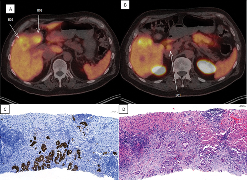 Fig. 1
Peer Reviews
No public reviews on file for this paper yet. If you reviewed it on a platform where reviews are public (OpenReview, ICLR, NeurIPS, ICML), you can paste yours below so the community can read it here.
Videos
No videos yet. Explain this paper in a talk, walkthrough, or lecture? Add one.
Taxonomy
TopicsCholangiocarcinoma and Gallbladder Cancer Studies · Prostate Cancer Treatment and Research · Peptidase Inhibition and Analysis
Introduction
Cholangiocarcinoma is an aggressive malignancy arising from the biliary tract epithelium with rising incidence and mortality. 1 Imaging commonly used for diagnostic workup include computed tomography (CT) scan and magnetic resonance imaging (MRI). Fluorine-18 (F-18) fluorodeoxyglucose positron emission tomography (FDG PET) scans can be used for investigating equivocal findings on radiographic imaging. Prostate-specific membrane antigen (PSMA) is known to be highly expressed in prostate adenocarcinoma allowing it to be leveraged as a target for both imaging and radioligand therapy in prostate cancer. However, PSMA is also commonly expressed in other malignancies such as breast cancer, thyroid carcinomas, and head and neck malignancies, which increases the chances of their incidental diagnosis on PSMA PET scans. 2 3 4 However, biliary tract cancers are not commonly expected to be incidentally diagnosed on PSMA PET imaging. Here, we describe the case of a patient with known prostate adenocarcinoma who was later incidentally diagnosed with advanced intrahepatic cholangiocarcinoma on PSMA PET/CT.
Case Report
A 66-year-old man with past medical history of prostate cancer, thyroid cancer, hypertension, and atrial fibrillation underwent F-18 PYL PSMA PET/CT for staging of localized prostate adenocarcinoma prior to scheduled radical prostatectomy. The F-18 PYL PSMA PET/CT showed a large area of heterogenous PYL uptake (SUV max = 16.9) involving liver segments 4B and 5 that was suspicious for a primary liver malignancy ( Fig. 1A ). Follow-up MRI revealed a 5.8 × 4.5 cm segment 4B/5 hepatic mass with adjacent gallbladder invasion and enlarged portacaval lymph node suggestive of metastatic disease. Ultrasound-guided needle biopsy of the liver was noted to be consistent with a well-differentiated adenocarcinoma. Immunostaining was positive for CK7 and negative for HepPar 1, NKX3.1, and TTF-1, favoring a diagnosis of cholangiocarcinoma ( Fig. 1C ). The patient underwent staging imaging with CT of the chest, abdomen, and pelvis, which revealed metastatic disease involving the anterior abdominal wall. The patient was treated for metastatic intrahepatic cholangiocarcinoma with standard of care chemoimmunotherapy (gemcitabine, cisplatin, and durvalumab). He remains in follow-up on maintenance immunotherapy.
Fluorine-18 (F-18) PYL prostate-specific membrane antigen positron emission tomography/computed tomography cross-sectional image showing ( A ) tracer-avid lesions in segment 4B/5 suggestive of primary malignancy (B02, B03) and ( B ) tracer-avid lesion indicative of metastatic spread to portocaval lymph node (B01). Histopathological examination demonstrating ( C ) focus of strong CK7 positivity on immunohistochemistry and ( D ) hematoxylin and eosin stain demonstrating well-differentiated adenocarcinoma.
Discussion
In the current report, we present a case of intrahepatic cholangiocarcinoma found incidentally on a F-18 PYL PSMA PET scan. As discussed earlier, cholangiocarcinoma is not typically expected to be incidentally diagnosed on a PSMA PET/CT.
On literature review, we found previous studies noting a high rate of positivity for PSMA on immunohistochemistry in cholangiocarcinoma. An analysis of 203 samples of cholangiocarcinoma found that 79.3% of samples were PSMA positive on immunohistochemistry. In addition, PSMA expression positively correlated with the grade and stage of cholangiocarcinoma. The PSMA positive cases were seen more in stages III and IV and high-grade disease compared with early-stage and low-grade disease, respectively. 5 A few prior case reports of intrahepatic cholangiocarcinoma detected PSMA PET imaging have been reported. 6 7 8 There are insufficient data to determine if PSMA PET can be reliably used to differentiate between metastases from prostate cancer and primary hepatobiliary malignancies. However, there should be a high index of suspicion for the latter, especially in the presence of multiple risk factors that increase the risk of primary hepatobiliary malignancies.
The mechanism of PSMA expression tends to differ between malignant tumors such as primary hepatobiliary malignancies and benign liver lesions. PSMA is primarily expressed on neovascular endothelium as opposed to increased expression by virtue of increased blood flow or folate receptors in macrophages in the case of benign liver lesions. 5 Interestingly, a recent analysis of 72 liver tumors also identified perivascular PSMA as a potential biomarker for distinguishing between primary cholangiocarcinoma and metastatic pancreatic adenocarcinoma, two diagnoses that are commonly difficult to distinguish by histology alone. 9 Altogether, these results suggest that PSMA PET merits further investigation for potential application in the diagnosis of unclear cases of cholangiocarcinoma. Furthermore, these findings may further serve as the basis for investigating the role of theranostic applications of PSMA expression in cholangiocarcinoma.
The reference list from the paper itself. Each links out to its DOI / PubMed record.
- 1Gad M M Saad A M Faisaluddin M Epidemiology of cholangiocarcinoma; United States incidence and mortality trends Clin Res Hepatol Gastroenterol 2020440688589332359831 10.1016/j.clinre.2020.03.024 · doi ↗ · pubmed ↗
- 2Bertagna F Albano D Giovanella L 68 Ga-PSMA PET thyroid incidentalomas Hormones (Athens)2019180214514930989578 10.1007/s 42000-019-00106-8 · doi ↗ · pubmed ↗
- 3Zhou W Halder S Herwald S Frequent amplification and overexpression of PSMA in basallike breast cancer from analysis of The Cancer Genome Atlas J Nucl Med 202465071004100638664014 10.2967/jnumed.123.266659 · doi ↗ · pubmed ↗
- 4Lawhn-Heath C Flavell R R Glastonbury C Hope T A Behr S C Incidental detection of head and neck squamous cell carcinoma on 68 Ga-PSMA-11 PET/CT Clin Nucl Med 20174204 e 218e 22028166149 10.1097/RLU.0000000000001569 · doi ↗ · pubmed ↗
- 5Chen L X Zou S J Li D Prostate-specific membrane antigen expression in hepatocellular carcinoma, cholangiocarcinoma, and liver cirrhosis World J Gastroenterol 202026487664767833505143 10.3748/wjg.v 26.i 48.7664 PMC 7789058 · doi ↗ · pubmed ↗
- 6Kang C Jiang J Y Lee M E Shen L Mansberg R Incidental intrahepatic hepatocellular cholangiocarcinoma detected on 68 Ga-PSMA PET/CT Clin Nucl Med 20224703 e 291e 29335020661 10.1097/RLU.0000000000003992 · doi ↗ · pubmed ↗
- 7Veenstra M MK Vegt E Segbers M Intra-arterial PSMA injection using hepatic arterial infusion pump in intrahepatic cholangiocarcinoma: a proof-of-concept study Eur Radiol Exp 20248019039090480 10.1186/s 41747-024-00496-4PMC 11294287 · doi ↗ · pubmed ↗
- 8Sun Y Wang H Yang Y You Z Zhao J Intrahepatic cholangiocarcinoma detected on 18 F-PSMA-1007 PET/MR imaging in a prostate cancer patient: a case report and literature review Front Oncol 2024141.408453 E 610.3389/fonc.2024.1408453 PMC 1119951838933442 · doi ↗ · pubmed ↗