The value of high-pitch scanning with Sn100kV and ADMIRE in CT examination of tuberculous destroyed lung: Identifying the optimal combination for ultra-low-dose imaging
Dong Jiang, Lixin Qin, Wenyang Pan, Shixiang Yan

TL;DR
This study shows that high-pitch CT scanning with Sn100kV and ADMIRE can produce good quality images for lung exams in tuberculosis patients while reducing radiation exposure and motion artifacts.
Contribution
The study introduces a novel ultra-low-dose CT technique using high-pitch scanning and energy spectrum purification for diagnosing tuberculous lung damage.
Findings
Group B (high-pitch scanning) had significantly fewer motion artifacts and 76.24% lower radiation dose compared to Group A.
Image quality scores in both groups met clinical standards, with no significant difference in lesion detection rates.
Group B showed better SNR and CNR in pulmonary window images despite higher noise levels.
Abstract
To investigate the application value of high-pitch scanning combined with energy spectrum purification using Sn100kV and ADMIRE in CT examinations of patients with tuberculous destroyed lung. A total of 60 patients with sputum mycobacterium tuberculosis smear positive and diagnosed with tuberculous lung damage on imaging were prospectively collected. The first CT examination utilized a conventional scanning mode with a fixed tube voltage of 120kV, CARE Dose4D activated, reference tube current set at 70mAs, and a pitch of 1.5. The interval between the initial and follow-up CT was over three months. During the follow-up CT, a high-pitch scanning mode combined with energy spectrum purification was employed, with a fixed tube voltage of Sn100kV, CARE Dose4D activated, reference tube current set at 300mAs, and a pitch of 3.2. The remaining parameters were consistent between the two CT…
Genes, proteins, chemicals, diseases, species, mutations and cell lines named across the full text — each resolved to its canonical identifier and authoritative record.
Click any figure to enlarge with its caption.
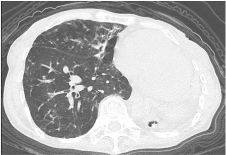 Figure 1
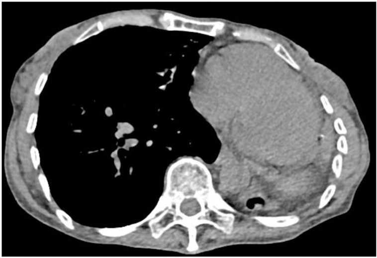 Figure 2
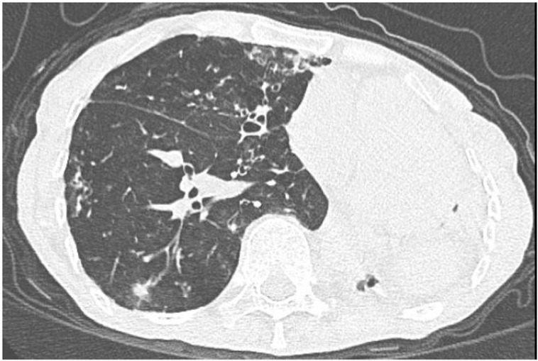 Figure 3
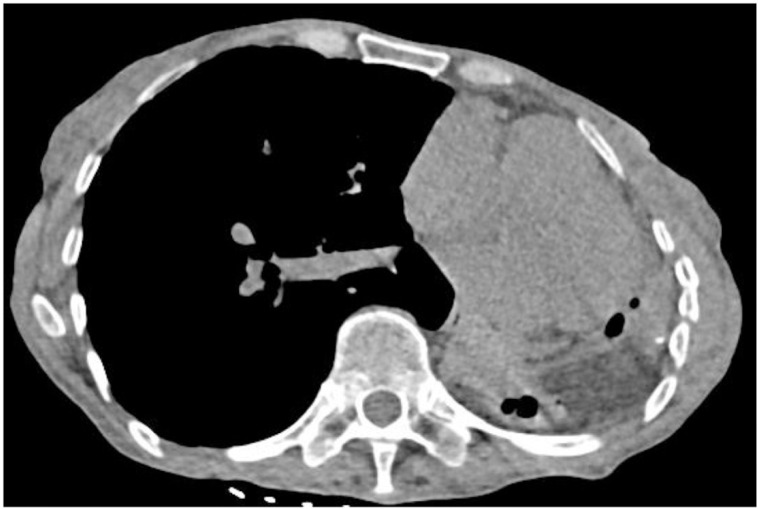 Figure 4
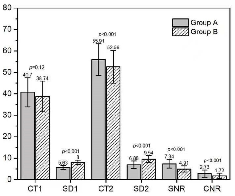 Figure 5
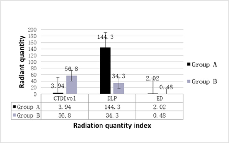 Figure 6
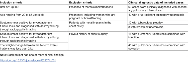 Figure 7
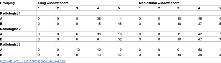 Figure 8
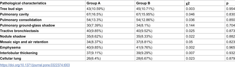 Figure 9
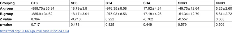 Figure 10
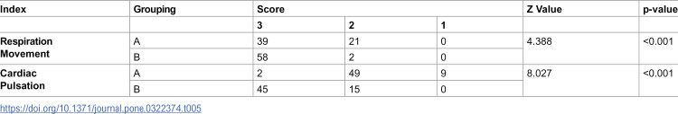 Figure 11
Peer Reviews
No public reviews on file for this paper yet. If you reviewed it on a platform where reviews are public (OpenReview, ICLR, NeurIPS, ICML), you can paste yours below so the community can read it here.
Videos
No videos yet. Explain this paper in a talk, walkthrough, or lecture? Add one.
Taxonomy
TopicsAdvanced X-ray and CT Imaging · Radiation Dose and Imaging · Medical Imaging Techniques and Applications