A genome-wide CRISPR screen identifies GRA38 as a key regulator of lipid homeostasis during Toxoplasma gondii adaptation to lipid-rich conditions
Mebratu A. Bitew, Tatiana C. Paredes-Santos, Parag Maru, Shruthi Krishnamurthy, Yifan Wang, Lamba O. Sangaré, Samuel Duley, Yoshiki Yamaryo-Botté, Cyrille Botte, Jeroen P.J. Saeij

TL;DR
A study finds that GRA38 helps the parasite Toxoplasma gondii manage lipids, which is crucial for its survival and virulence.
Contribution
Identifies GRA38 as a novel lipid homeostasis regulator in Toxoplasma gondii using a CRISPR screen.
Findings
GRA38 has phosphatidic acid phosphatase activity, essential for lipid homeostasis in Toxoplasma.
Disruption of GRA38 causes lipid imbalances and reduced parasite virulence in mice.
GRA38's activity is linked to adaptation to lipid-rich environments.
Abstract
Intracellular parasites like Toxoplasma gondii scavenge host nutrients, particularly lipids, to support their growth and survival. Although Toxoplasma is known to adjust its metabolism based on nutrient availability, the mechanisms that mediate lipid sensing and metabolic adaptation remain poorly understood. Here, we performed a genome-wide CRISPR screen under lipid-rich (10% Fetal Bovine Serum (FBS)) and lipid-limited (1% FBS) conditions to identify genes critical for lipid-responsive fitness. We identified the Toxoplasma protein GRA38 as a lipid-dependent regulator of parasite fitness. GRA38 exhibits phosphatidic acid (PA) phosphatase (PAP) activity in vitro, which is significantly reduced by mutation of its conserved DxDxT/V catalytic motif. Disruption of GRA38 led to the accumulation of PA species and widespread alterations in lipid composition, consistent with impaired PAP…
Genes, proteins, chemicals, diseases, species, mutations and cell lines named across the full text — each resolved to its canonical identifier and authoritative record.
Click any figure to enlarge with its caption.
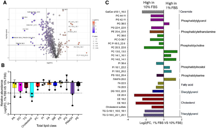 Figure 1
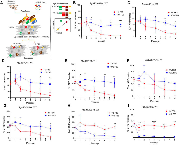 Figure 2
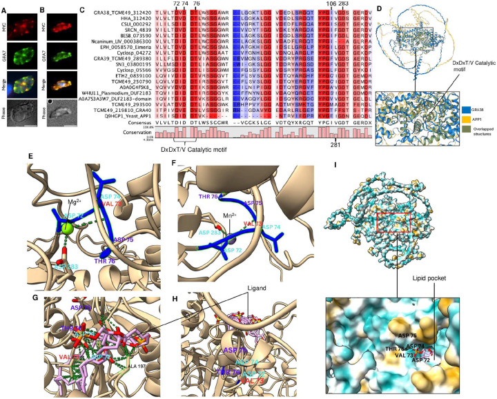 Figure 3
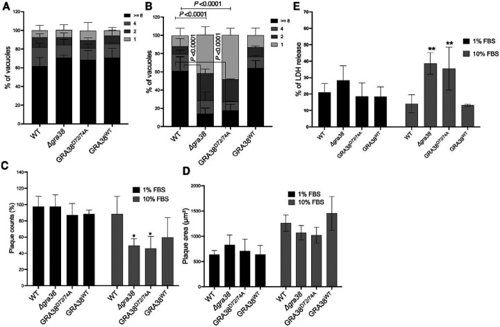 Figure 4
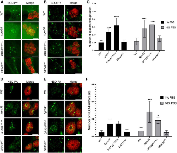 Figure 5
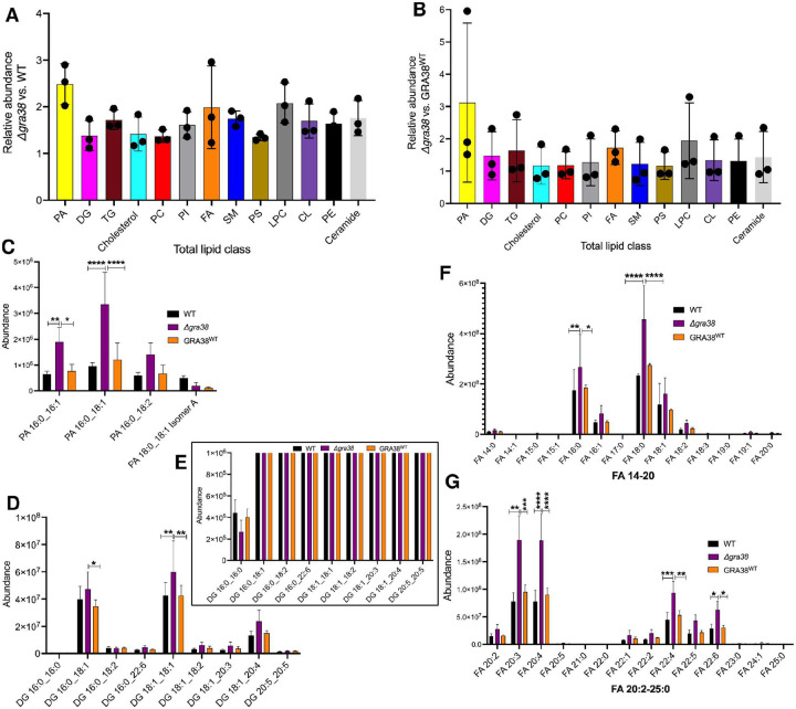 Figure 6
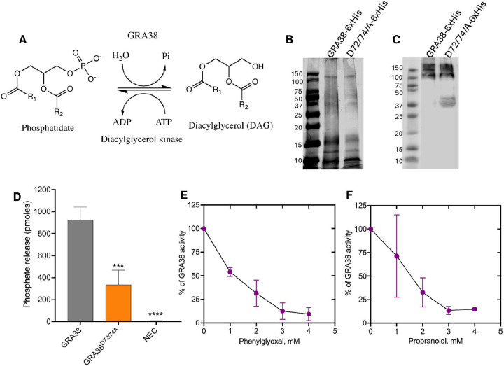 Figure 7
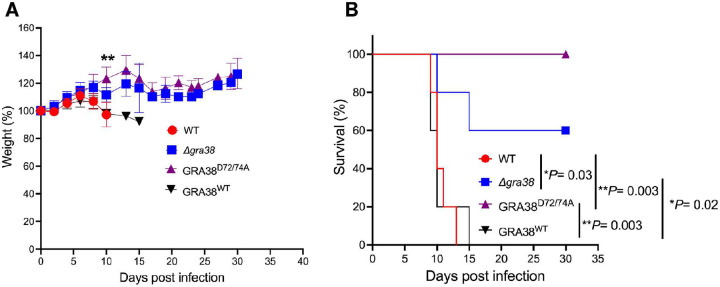 Figure 8
Peer Reviews
No public reviews on file for this paper yet. If you reviewed it on a platform where reviews are public (OpenReview, ICLR, NeurIPS, ICML), you can paste yours below so the community can read it here.
Videos
No videos yet. Explain this paper in a talk, walkthrough, or lecture? Add one.
Taxonomy
TopicsToxoplasma gondii Research Studies · Autophagy in Disease and Therapy · Cytomegalovirus and herpesvirus research
Introduction
Toxoplasma gondii is an obligate intracellular parasite that causes toxoplasmosis, a disease with severe consequences in immunocompromised individuals and during pregnancy (1). To sustain its replication and survival, Toxoplasma relies extensively on host-derived nutrients, especially lipids. Its ability to infect virtually any nucleated cell of warm-blooded animals highlights a remarkable metabolic flexibility, allowing it to adapt to diverse cellular environments.
Within host cells, Toxoplasma resides in the parasitophorous vacuole (PV), a specialized compartment that serves as the primary interface for host-parasite interactions and nutrient acquisition (2). Nutrient availability, particularly lipids, strongly influences parasite growth, development, and life-stage differentiation, highlighting the significance of host metabolic status in Toxoplasma biology.
To satisfy its lipid requirements, Toxoplasma uses a dual strategy of de novo synthesis and extensive host lipid scavenging. While the parasite can synthesize fatty acids in the apicoplast via the FASII pathway, it cannot produce cholesterol de novo and must scavenge it from the host (2–12). To scavenge host-derived lipids, Toxoplasma actively recruits host organelles, including the endoplasmic reticulum (ER), Golgi apparatus, mitochondria, and giant multivesicular bodies (gMVBs) arising from host organelles, to the PVM. It also hijacks Rab-dependent host vesicle trafficking pathways (10,13,14). The parasite subsequently internalizes these lipid-containing structures into the PV, facilitated by the Endosomal Sorting Complex Required for Transport (ESCRT) machinery (15,16) and enzymes such as TgLCAT, a phospholipase A2 responsible for releasing lipids for parasite uptake (17).
Once inside the PV, these lipids are processed and trafficked by parasite-encoded proteins, including the ATP-binding cassette (ABC) G transporter TgABCG107 (18); TgHAD-2SCP-2, which contains sterol carrier protein-2 [SCP-2] domains; TgACBP1, an acyl-CoA binding protein (19); and TgNCR1, a Niemann-Pick Type C1-related transporter (20). Together, these components orchestrate lipid uptake, remodeling, and storage to support parasite replication and prevent lipotoxicity.
This capacity to balance de novo lipid synthesis and host lipid scavenging enables Toxoplasma to thrive in host cells with vastly different lipid profiles—from lipid-rich macrophages to tissues like liver or brain (21). However, the molecular mechanisms underlying the parasite’s ability to sense lipid availability and mediate metabolic adaptation remain poorly defined.
Prior work has shown that Toxoplasma modulates its lipid acquisition strategies based on nutrient availability. For example, under lipid-limited conditions (e.g., 1% Fetal Bovine Serum (FBS)), the parasite upregulates its apicoplast-based fatty acid synthesis pathway (FASII) and lipid scavenging mechanisms (6,9,10,12,13,22). In contrast, under lipid-rich conditions (e.g., 10% FBS), scavenging predominates while de novo synthesis is down regulated. Under these conditions, scavenged lipids are primarily channeled into parasite lipid droplets (LD), which are mobilized during parasite division to prevent lipotoxicity (9,23). This adaptive response highlights how the essentiality of enzymes in Toxoplasma is context-dependent.
This adaptive flexibility is not unique to Toxoplasma. In Plasmodium falciparum, the malaria-causing parasite, the apicoplast FASII pathway is dispensable under normal blood stage conditions but becomes essential during lipid starvation (6,24). Likewise, Toxoplasma lipid-modifying enzymes, such as acyl-CoA synthetases TgACS1 and TgACS3, the acyltransferase TgATS2, and the phosphatidic acid phosphatase (PAP) TgLIPIN, demonstrate nutrient-dependent importance, highlighting their potential as drug targets under specific metabolic conditions (6,9,22,25,26).
Importantly, existing genome-wide CRISPR-Cas9 screens have primarily been performed under nutrient-rich conditions (10% FBS), potentially missing genes important for adaptation to variable host environments (56). To address this, we performed a genome-wide CRISPR screen comparing Toxoplasma fitness in lipid-limited (1% FBS) vs. lipid-rich (10% FBS) conditions to identify genes that mediate metabolic adaptation to lipid availability. Among the top hits was GRA38, a dense granule protein containing a Haloacid Dehalogenase (HAD) motif (27). We show that GRA38 functions as a PAP, is important for lipid homeostasis under lipid-rich conditions, and localizes to the PV lumen. Disruption of GRA38 leads to altered lipid profiles, with an accumulation of PA species and changes in diacylglycerol (DAG) species, premature egress under lipid-rich conditions, and reduced virulence in mice. Together, these findings establish GRA38 as a key mediator of lipid-responsive metabolic adaptation and pathogenesis in Toxoplasma gondii.
Results
Host lipidomic profiling under 1% and 10% FBS reveals distinct serum-dependent lipid alterations
Serum is the primary source of lipids in cell culture, and variations in FBS concentration are known to influence cellular lipid metabolism. To establish baseline differences in host lipid composition under these conditions, we performed lipidomic profiling of uninfected human foreskin fibroblasts (HFFs) cultured in media supplemented with either 1% or 10% FBS. Liquid chromatography-mass spectrometry (LC-MS) identified 856 lipid species, with 111 lipid species showing statistically significant differences between conditions (Table S1, Figure 1). Principal component analysis (PCA) and unsupervised clustering showed clear separation between 1% and 10% FBS samples, confirming the substantial impact of serum concentration on host lipid composition (Figure S1). The volcano plot (Figure 1A) highlights lipid species with significant changes in abundance.
Consistent with the clustering and PCA results, total lipid abundance was markedly higher in HFFs cultured in 10% FBS compared to 1% FBS, underscoring the lipid-rich nature of the high-serum condition (Figure 1B). Several lipid classes showed substantial enrichment in 10% FBS, including phosphatidic acid (PA), diacylglycerol (DG), triacylglycerol (TG), and cholesterol. Sphingomyelins (SM), and a range of phospholipids—such as phosphatidylcholine (PC), phosphatidylethanolamine (PE), and phosphatidylmethanol (PMeOH)—were also elevated under high-serum conditions (Figure 1B). In contrast, the abundance of phosphatidylinositol (PI), phosphatidylserine (PS), Fatty acids (FA) and phosphatidylglycerol (PG) remained relatively unchanged between conditions, (Figure 1B), suggesting selective regulation of specific phospholipid species in response to serum availability.
Figure 1C provides selected examples of individual lipid species that are differentially regulated.
Among the lipid species that changed between conditions, the following trends were observed. Lipid storage pathways were consistently enhanced under high-serum conditions. Several triacylglycerol species, including TG 18:2_18:2_18:2, TG O-18:0_20:1_20:1, and TG O-18:1_16:0_18:1, were significantly elevated in 10% FBS, consistent with increased incorporation of serum-derived unsaturated and ether-linked fatty acids into neutral lipid stores. Cholesterol derivatives—including cholesterol sulfate, cholesterol esters (CE 16:0, CE 18:2 and CE 20:4)—were also enriched, reflecting uptake of lipoprotein-associated sterols and intracellular esterification for storage. In addition, DG 16:0_22:6, DG 18:1_18:1 and DG 16:0_18:1 were elevated, consistent with increased lipid turnover and incorporation of serum-derived fatty acids, including long-chain polyunsaturated fatty acid (PUFAs) species. Fatty acid, FA 20:5, was also elevated in 10% FBS, consistent with increased oxidative lipid metabolism, while FAHFA 20:0 was reduced (Figure 1C), suggesting suppression of endogenous signaling lipid synthesis in nutrient-replete conditions. Phospholipids underwent acyl chain–specific remodeling in response to serum availability. In 10% FBS, there was consistent enrichment of PUFA-containing species across classes, PC 20:3_22:6, PC 38:6, PS 18:0_20:4, PI 38:4 as well as ether-linked forms such as PC O-38:7 (28,29). Phosphatidylethanolamine species followed this trend with PE 20:4_22:6 elevated in 10% FBS. These shifts are consistent with increased incorporation of serum-derived arachidonic, adrenic, and docosahexaenoic acids. In contrast, short-chain and saturated or monounsaturated species—including PC 14:0_14:1, PC 14:1_16:1, PC 13:0_13:0, PI 18:0_20:2, and PI 16:1_22:2 were more abundant in 1% FBS (Figure 1C), consistent with enhanced reliance on endogenously synthesized fatty acids and selective retention of energetically accessible lipid species under lipid-limiting conditions. Phosphatidylglycerol species, including PG 22:4_22:6, PG 42:11, and PG 44:11, were also elevated in 10% FBS, consistent with increased incorporation of polyunsaturated fatty acids into mitochondrial lipid pools whereas PG 38:5 was highly abundant in 1% FBS (Figure 1C). Sphingolipids were differentially regulated by serum availability. In 10% FBS several ceramide species—including Cer d16:1_16:0, Cer d18:1_16:0, Cer d18:2_20:0, and Cer d34:0—were elevated, along with GalCer d18:1_16:0, consistent with increased availability of sphingoid bases and saturated acyl-CoAs required for ceramide and glycosphingolipid biosynthesis (30). These changes are indicative of enhanced flux through sphingolipid metabolic pathways in lipid-replete conditions.
Together, these results confirm that host lipid profiles differ markedly at the lipid species level between 1% and 10% FBS conditions, validating this system for investigating Toxoplasma genes that differentially affect parasite fitness under lipid-rich vs. lipid-limiting environments using CRISPR-based screening.
Genome-wide CRISPR screen identifies Toxoplasma genes that determine parasite fitness at different serum concentrations
Toxoplasma has a remarkable ability to balance lipid acquisition, synthesis, and storage based on nutrient availability (2). To identify the genes involved in this metabolic adaptability, we performed a genome-wide CRISPR screen under both lipid-rich (10% serum) and lipid-limited (1% serum) conditions. A parasite population that had stably integrated a pooled guide RNA library was grown in HFFs under each condition and serially passaged, allowing parasites with condition-specific fitness defects to be outcompeted. At each passage, parasite genomic DNA was extracted and guide RNA abundance was quantified using next-generation sequencing (Figure 2A). We then ranked genes by differential guide RNA representation, applying a log2 fold-change cutoff and an adjusted p-value threshold to select the top candidate genes with significant fitness effects in 10% vs. 1% serum. To capture both immediate and long-term consequences of gene disruption under varying lipid conditions, we evaluated parasite fitness at both early passages (mean of P4/5) as well as at a late passage (P8). The top candidate genes with significant fitness effects in 10% vs. 1% serum are listed in Table 1, while additional significant hits are provided in Table S2.
Under lipid-limited conditions, genes involved in endogenous lipid processing –TGGT1_310150 (TgACS2), TGGT1_212130 (patatin-like phospholipase), and TGGT1_275590 (DGAT2L1, Table S2)– had significant fitness defects, showing the parasite’s reliance on intrinsic lipid synthesis and remodeling when exogenous lipids are scarce. In addition, genes related to RNA processing and stress responses, such as TGGT1_248110 (repressor of RNA polymerase III transcription, MAF1), TGGT1_231440 (LSm4, associated with U6 snRNA), TGGT1_269175 (Usb1, U6 snRNA phosphodiesterase), and TGGT1_306380 (U1 small nuclear ribonucleoprotein C (SNRPC), also known as U1-C zinc finger protein (Table S2)), had reduced fitness. Additional hits –TGGT1_212930 (NFU1 Fe-S cluster scaffold homolog), TGGT1_208090 (5-formyltetrahydrofolate cyclo-ligase), and TGGT1_320280 (orotidine 5’-monophosphate decarboxylase)– suggest that enhanced mitochondrial function and nucleotide biosynthesis also contribute to parasite survival under low serum conditions. Notably, four genes –GRA57, GRA70, GRA71 and TGGT1_200370 (encoding for the farnesyl transferase beta subunit)– previously identified as top hits in a screen for Toxoplasma fitness in IFNү-stimulated HFFs (32) also showed increased fitness defects in 1% serum conditions.
In contrast, under lipid-rich conditions, the top hits (Table 1) included genes important for managing lipid abundance. These consisted of TGGT1_242380 (fatty acid elongase), TGGT1_236660 (START domain-containing STARD3 homolog), which redistributes lipids, TGGT1_244270 (ABCG87 transporter), which exports lipids, TGGT1_254270 (TLCD4 orthologue), which is involved in membrane lipid remodeling, TGGT1_290980 (serine C-palmitoyltransferase), which synthesizes sphingolipid precursors, and TgACC2 (acetyl-CoA carboxylase)(Table S2), which initiates de novo fatty acid biosynthesis. Collectively, these genes act to prevent lipid overload and toxicity (33). Furthermore, genes related to vesicle trafficking –TGGT1_309610 (TMEM230 orthologue), TGGT1_310460 (Rab6), and TGGT1_237280 (TgTBC6) (34,35)– as well as the dense granule protein diacylglycerol kinase 2 (TgDGK2)(Table S2) (36) and GRA38 (a homologue of GRA39)(37), were identified as important for fitness in 10% serum. This suggests that the parasite requires more enzymatic activity to manage the increased lipid content in the host and additional mechanisms to acquire these resources. Taken together, our data suggest that the parasite senses host lipid content and rewires its metabolic program accordingly, making certain enzymes more essential or dispensable under different physiological and nutrient conditions.
Growth competition assay confirms differential fitness of selected knockouts in lipid-rich and lipid-limited conditions
To validate the genome-wide CRISPR screen results, growth competition assays were performed with selected knockouts under lipid-rich (10% FBS) and lipid-limited (1% FBS) conditions (Figure 2B–I). Knockout parasite strains were generated using a CRISPR/Cas9-based approach, where sgRNA targeting facilitated gene disruption via integration of a drug selection cassette, followed by clonal selection and PCR confirmation. Equal numbers of wild-type (WT) and knockout parasites were mixed, passaged in HFFs, and analyzed via plaque assays at passages 0, 1, 2, 4, 6, and 8 to assess fitness based on the knockout-to-total parasite ratio. Several knockouts, including Δ261400 (38), Δgra57, Δgra70, Δgra71, Δ200370, and Δ264740 (32), had fitness defects specifically under lipid-limited conditions (1% FBS), while maintaining WT-like fitness in lipid-rich environments. These knockouts showed progressively declining representation under 1% FBS, with Δgra70 and Δ264740 showing particularly pronounced reductions, while Δ200370 had a more moderate effect. In contrast, Δ269620 and Δgra38 had fitness defects under lipid-rich conditions (10% FBS). Δ269620 declined significantly in 10% FBS, but remained unaffected in 1% FBS, suggesting a role in lipid processing under lipid-abundant conditions. Δgra38 had the strongest defect in 10% FBS, with little impact in 1% FBS, indicating its fitness is specifically tied to lipid abundance. These results validate the CRISPR screen results and highlight the roles of the identified genes in parasite adaptation to lipid availability. GRA38 was prioritized for further investigation due to its strong phenotype and significant impact on parasite fitness in lipid-rich environments.
Structural predictions indicate that GRA38 is an enzyme within the Haloacid Dehydrogenase (HAD) superfamily.
To determine the localization of TGGT1_312420 (GRA38) in parasites, we introduced the C-terminal MYC tag in the endogenous GRA38 locus. In intracellular parasites, GRA38 is localized to the PV lumen along with GRA7 (Figure 3A). In extracellular parasites (Figure 3B), GRA38 colocalized with GRA7 within dense granules. Blast analysis and alignment of orthologous proteins revealed that GRA38 is highly conserved among apicomplexan parasites (Figure 3C). Although GRA38 is currently annotated as a hypothetical protein with no known function, it contains a conserved putative catalytic DxDx(T/V)(L/V) motif, characteristic of haloacid dehalogenase (HAD) enzymes (27,39). We next used Alphafold (40) and Foldseek (41) to identify proteins with structural similarities. Significant matches (E-values < 0.05) were found with actin patch proteins (APP1)(Uniprot P53933, Q9HGP1), which contain the phosphatidate phosphatase catalytic domain (42). Similar results were observed when analyzing GRA39 or when focusing on the first 289 amino acids of GRA38 (e.g., similarity to phosphatidate phosphatase APP1 catalytic domain-containing proteins from Eutreptiella gymnastica (A0A7S4CYL7) and Vitrella brassicaformis (A0A0G4FSK8). Pairwise structural alignment of both GRA38 and APP1 using FoldMason in Foldseek (43) showed structural similarities, especially around the DxDxT/V motif (an MSA LDDT score of 0.646) (Figure 3D).
HAD enzymes are known to coordinate divalent metal ions such as Mg^2+^ or Mn^2+^ to facilitate catalytic activity (44). Structural modeling using AlphaFold predicted that both Mg^2+^ and Mn^2+^ ions can bind to GRA38 specifically to Asp72, Val73 and Asp283 (Figure 3E/F), aligning with the classical Mg^2+^ and Mn^2+^ dependent mechanism of action observed in PAPs, such as lipin/Pah superfamily, within the HAD family (45).
Next, we docked PA and a non-substrate lipid, cholesterol, into the predicted binding site of GRA38 using the SwissDock platform, powered by AutoDock Vina for molecular docking (46,47). PA demonstrated a strong binding affinity to the hydrophobic pocket formed by GRA38 (Figure 3I), positioning itself in close proximity to the DxDxT/V catalytic motif (Figure 3G). Specifically, PA was observed to directly interact with residues in the DxDxT/V motif, consistent with a role in catalysis (Figure 3G). In contrast, docking cholesterol, which lacks the same polar head group and charge distribution as PA, revealed no specific interactions within the catalytic pocket. Instead, cholesterol remained positioned outside of the binding site, failing to engage with the DxDxT/V motif (Figure 3H).
GRA39, a homologue of GRA38 (37), similarly possesses a conserved DxDx(T/V)(L/V) motif and shares structural features with PA phosphatases. Modeling suggests that Mg2+ and Mn2+ ions are coordinated by Asp98, Asp100, and Asp257 (Figure S2). Like GRA38, PA exhibits strong binding affinity to GRA39’s hydrophobic pocket near the catalytic motif, while cholesterol remains external (Figures S2C/D). We therefore hypothesized that GRA38 has PAP activity and plays a role in regulating lipid homeostasis and mobilization within the PV lumen.
The DxDxT/V motif is required for the catalytic function of GRA38 in lipid-rich conditions
To investigate the role of the DxDxT/V motif in GRA38’s function, the DxDxT/V motif was mutated to AxAxT/V (D72/74A), and parasites were complemented with plasmids encoding WT or GRA38^D72/74A^ versions of GRA38 (Figure S4). The replication of WT, Δgra38, GRA38^WT^, and GRA38^D72/74A^ parasites was compared in HFFs cultured with 1% and 10% FBS media. Parasite replication was assessed by counting the number of parasites per vacuole after 24 hours of infection (Figure 4A). In media containing 1% FBS, Δgra38 parasites exhibited normal parasite replication compared to WT parasites (Figure 4A). However, when cultured in media containing 10% FBS, Δgra38 parasites showed a significant reduction in vacuoles containing eight parasites and a higher percentage of vacuoles containing one parasite, compared to the vacuoles of WT parasites. This indicates that the growth rate of Δgra38 parasites is slower or that they might egress earlier. Complementation of Δgra38 parasites with a WT copy of GRA38 rescued this phenotype (Figure 4A), while complementation with the GRA38^D72/74A^ did not (Figure 4A), indicating a severe growth defect under lipid-rich conditions. This highlights the importance of the DxDxT/V motif for parasite growth in a high-lipid environment.
To further investigate the role of the DxDxT/V motif in GRA38 function, plaque assays were performed using the same parasite strains. Plaque counts and areas were measured five days post-infection in HFFs cultured with 1% or 10% FBS media (Figure 4B, C). The area (Figure 4C) and number of plaques (Figure 4D) formed by Δgra38 and GRA38^D72/74A^ parasites under 1% FBS conditions were not significantly different from WT parasites. However, in 10% FBS media, the relative plaque counts of Δgra38 and GRA38^D72/74A^ parasites were significantly lower than those of WT or GRA38^WT^ parasites (Figure 4B). The plaques formed by Δgra38 and GRA38^D72/74A^ parasites are slightly smaller than those formed by WT or GRA38^WT^ parasites, but this was not statistically significant (Figure 4C). These results indicate that the DxDxT/V motif is essential for GRA38’s role in supporting parasite replication and survival in lipid-rich environments.
Deletion of GRA38 leads to host cell death consistent with early parasite egress in lipid rich environment
We observed a difference in the number of parasites per vacuole between strains grown in media with 1% versus 10% FBS (Figure 4A), while the plaque areas remained comparable across these conditions (Figure 4C). This led us to hypothesize that the Δgra38 and GRA38^D72/74A^ strains may undergo early egress. To test this, we measured host cytoplasmic lactate dehydrogenase (LDH) release, which indicates host cell death (48). We observed no difference in host cell death in HFFs infected with parasites grown in 1% FBS (Figure 4D). However, there was a significant increase in cell death in HFFs infected with Δgra38 and GRA38^D72/74A^ strains grown in 10% FBS, consistent with parasite egress (Figure 4D).
GRA38 regulates lipid accumulation and phosphatidic acid homeostasis in Toxoplasma
To investigate the role of GRA38 in lipid mobilization and homeostasis, we infected host cells with different parasite strains in media containing either 1% or 10% FBS and compared the number of parasite lipid droplets (LD) using BODIPY 493/503 solution. Quantification of parasite LD revealed a 135% and 139% increase in LD accumulation in Δgra38 parasites compared to WT or GRA38^WT^ parasites when grown in 1% FBS and 10% FBS, respectively (Figure 5A–C). Similarly, the GRA38^D72/74A^ strain accumulated 220% and 182% more lipid droplets than WT or GRA38^WT^ parasites in 1% and 10% FBS, respectively (Figure 5A–C). These results indicate that the absence of GRA38 disrupts LD content, potentially affecting neutral lipid accumulation, such as DAG, TG. This would be compatible and logical with the putative function of GRA38 as a PA phosphatase (PAP) as previously reported for TgLipin, a parasite PAP, whose disruption affects LD content, and parasite survival (9). This further points at the role of GRA38 in the regulation of lipid acquisition and metabolism during Toxoplasma infection.
To determine whether GRA38 plays a role in PA metabolism, HFFs were infected with different strains of Toxoplasma for 24 hours and then further incubated with nitro-benzoxadiazole (NBD)-conjugated PA for six additional hours. Fluorescence imaging of the probe-labeled parasites revealed the uptake of NBD-PA, evident as punctate intracellular NBD droplets distributed throughout the PV lumen and within the parasite body (Figure 5D–F). Δgra38 knockout parasites had significantly higher levels of NBD-PA compared to WT and complemented strains (Figure 5D–F). Similarly, GRA38^D72/74A^ knockout parasites had an increase in PA levels (Figure 5D–F), suggesting that the DxDxT/V motif in GRA38 is important for its catalytic role in converting PA to DAG. Additionally, both Δgra38 and GRA38^D72/74A^ parasites had slightly higher NBD-PA levels when grown in 10% FBS compared to 1% FBS, most likely because of the high nutrient content at 10% FBS, which favors host lipid scavenging by the parasite (6, 9), therefore potentially exacerbating PA accumulation.
Disruption of GRA38 alters phosphatidic acid metabolism and lipid profiles in Toxoplasma
To assess how GRA38 disruption affects lipid metabolism in Toxoplasma, we performed lipidomic profiling using liquid chromatography-mass spectrometry (LC-MS) on Δgra38, WT, and complemented (GRA38^WT^) strains grown in HFFs with 10% FBS, a lipid-rich condition where GRA38 contributes to parasite fitness. A total of 734 lipid species were identified by comparing retention times and mass spectra to in-house standards, generating robust profiles across three biological replicates. PCA and hierarchical clustering (Figure S5) confirmed distinct and reproducible lipidomic shifts between strains. Differential lipid abundance was further assessed using direct comparisons across strains (Figure 6).
Δgra38 parasites showed increased total lipid abundance compared to WT and GRA38^WT^ strains (Figure 6A, B), consistent with disrupted lipid homeostasis. Notably, cholesterol, TG, SM, ceramides, and several phospholipids—including lysophosphatidylcholine (LPC), phosphatidylethanolamine (PE), phosphatidylinositol (PI), phosphatidylcholine (PC), and cardiolipin (CL)—were elevated in Δgra38 parasites. Given the predicted role of GRA38 as a PAP converting phosphatidic acid (PA) to diacylglycerol (DAG), we examined PA species in detail (Figure 6C). Several PA species, including PA 16:0_16:1, PA 16:0_18:1, and PA 16:0_18:2 were more abundant in Δgra38 parasites, consistent with impaired PA hydrolysis due to loss of PAP activity. PA levels were restored to WT levels in the GRA38^WT^ strain, confirming complementation.
DAG species, the expected products of PAP activity, showed a more complex pattern (Figure 6D, E). For example, DAG 16:0_16:0 was reduced in Δgra38 parasites, consistent with impaired PA-to-DAG conversion. However, other DAGs such as DG 16:0_18:2, DG 16:0_22:6, DG 18:1_18:2, DG 18:1_20:3 and DG 20:5_20:5 remained unchanged (Figure 6D) and certain DAGs, including DG 16:0_18:1 and DG 18:1_18:1, were elevated in Δgra38 parasites (Figure 6D), suggesting selective effects on DAG metabolism or compensation through alternate pathways. DAG levels in the GRA38^WT^ strain were comparable to WT, supporting GRA38’s role in PA-to-DAG conversion.
Fatty acid (FA) profiles reflected downstream effects of disrupted lipid metabolism (Figure 6F, G). Δgra38 parasites showed distinct changes in both medium-chain FAs (e.g., FA 16:0, FA 16:1, FA 18:0, FA 18:1, FA 18:2) and long-chain polyunsaturated FAs (e.g., FA 20:2, FA 20:3, FA 22:1, FA 22:2, FA 22:4, FA 22:5, and FA 22:6). Notably, FA 20:4 (arachidonic acid), a host-derived PUFA scavenged by Toxoplasma, was elevated in Δgra38 parasites. FA profiles in GRA38^WT^ parasites were restored to WT levels, further confirming the metabolic impact of GRA38 disruption. Together, these data demonstrate that loss of GRA38 significantly alters the parasite’s lipid composition, leading to increased PA, selective changes in DAG species, and broad remodeling of phospholipid and fatty acid pools.
GRA38 exhibits PAP activity in vitro, which is significantly reduced by mutation of key catalytic residues
To determine whether GRA38 possesses PAP activity, recombinant GRA38–6xHis was expressed in and purified from Escherichia coli BL21 cells. The enzymatic activity of GRA38 was assessed using a colorimetric malachite green phosphate assay, which quantifies the release of free inorganic phosphate (Pi) during the conversion of PA to DAG (Figure 7A–C).
Reactions containing GRA38–6xHis yielded an average release of 925 pmol of free phosphate, confirming its PAP activity (Figure 7D). In contrast, the non-enzyme control showed negligible phosphate release (~0 pmol). To assess the contribution of conserved catalytic residues to PAP activity, site-directed mutagenesis of GRA38 was performed to generate the GRA38^D72/74A^ mutant. The purified GRA38^D72/74A^-6xHis protein exhibited significantly reduced activity, releasing only 335 pmol of phosphate, a marked reduction compared to wild-type GRA38–6xHis (Figure 7D). These results indicate that D72 and D74 are critical for GRA38’s PAP activity.
To further validate GRA38’s PAP activity and assess its susceptibility to enzymatic inhibition, a dose-dependent inhibition assay was performed using phenylglyoxal and propranolol, two known PAP inhibitors (49). GRA38–6xHis was incubated with increasing concentrations (0–4 mM) of each inhibitor and phosphate release was quantified. Both inhibitors suppressed PAP activity in a dose-dependent manner. At 1 mM, propranolol reduced phosphate release by approximately 29% (Figure 7F), while phenylglyoxal inhibited activity by 45% (Figure 7E). At 2–3 mM, inhibition progressively increased, and at 4 mM, phosphate release was reduced by 85–90% (Figures 7E and 7F). These findings confirm that GRA38 is a functional PAP enzyme whose activity depends on key catalytic residues and can be pharmacologically inhibited, supporting its potential role in lipid metabolism and Toxoplasma intracellular replication.
GRA38 plays a role in Toxoplasma virulence
To evaluate the role of GRA38 in the in vivo virulence of Toxoplasma, we performed intraperitoneal infections in CD-1 mice using 100 tachyzoites from different parasite strains: WT (RH Cas9 Luc+ Δhxgprt), RH Cas9 Δgra38, RH Cas9 GRA38^WT^, and RH Cas9 GRA38^D72/74A^. Mice infected with WT parasites showed significant weight loss (Figure 8A) and a decline in overall health throughout the experiment. None of the mice infected with WT parasites survived the full duration of the study (Figure 8B). Similarly, all mice infected with GRA38^WT^ parasites succumbed during the experiment (Figure 8B). In contrast, three out of five mice infected with Δgra38 parasites survived (Figure 8B). Remarkably, all mice infected with GRA38^D72/74A^ parasites survived the entire experiment (Figure 8B). Surviving mice were monitored for signs of infection, including rough fur and lethargy, and all tested positive for Toxoplasma-specific antibodies, confirming successful infection. These results demonstrate that GRA38 plays a role in the virulence of Toxoplasma.
Discussion
Our study demonstrates that GRA38 functions as a PAP in Toxoplasma, highlighting its important role in balancing PA and diacylglycerol (DAG) within the PV. Maintaining this PA-DAG equilibrium appears important for parasite lipid homeostasis and influences parasite replication, egress, and overall survival under lipid-rich conditions. High sequence conservation of GRA38 among apicomplexan parasites suggests that its lipid regulatory function may be evolutionarily conserved and likely plays a role in adapting to nutrient fluctuations within the PV. Thus, our findings position GRA38 as a newly recognized PAP with a non-redundant role in safeguarding the parasite from PA-driven lipotoxic stress, especially when lipids are abundant.
GRA38’s DxDxT/V catalytic motif, characteristic of the HAD superfamily (27,39) and PAPs (9,27,39,45,50–53), is predicted to facilitate the conversion of PA into DAG, a key metabolic step linking lipid scavenging to membrane biosynthesis and signaling. Disruption of GRA38 caused accumulation of multiple PA species, indicating impaired turnover, but also led to increased levels of several DAGs, particularly those enriched in PUFAs. This pattern suggests that PA-to-DAG conversion is not completely abolished in the Δgra38 mutant, but is instead altered in specificity or efficiency. These findings point to a selective perturbation of the PA-to-DAG axis, rather than a uniform block, and imply the existence of compensatory or partially redundant enzymes that maintain some level of DAG production in the absence of GRA38. One likely candidate for such compensation is GRA39, a GRA38 homologue sharing structural features and the conserved DxDx(T/V)(L/V) motif. This could explain the selective buildup of certain PA and DAG species observed in the lipidomic data. More broadly, the accumulation of PUFA-rich DAGs, phospholipids, lysophospholipids, and triacylglycerols suggests that without GRA38, Toxoplasma struggles to efficiently remodel host-derived lipid cargo, resulting in bottlenecks in lipid metabolism and disrupted homeostasis. These effects are particularly striking under lipid-rich conditions, where Δgra38 parasites exhibit impaired replication and premature egress. Elevated PA levels may activate inappropriate signaling cascades (59), shortening the replication phase and triggering early egress from host cells. Although the mutant shows minimal defects under lipid-poor conditions, the imbalance between PA and DAG likely becomes more pronounced as exogenous lipid availability increases. This highlights GRA38’s essential role in buffering the parasite against lipotoxic stress during nutrient-rich growth.
In sum, GRA38 appears to function as a key integrator of host lipid salvage within the parasitophorous vacuole, directing incoming lipids into productive biosynthetic routes and preventing the accumulation of poorly processed intermediates. Its activity is especially critical when lipid supply is abundant, where precise control over PA metabolism helps the parasite balance membrane synthesis, storage, and replication timing. Future studies examining GRA38 and GRA39 in combination—through double mutants or comparative lipidomics—will be necessary to clarify the division of labor between these PV-localized PAPs and to fully define their contributions to the regulation of Toxoplasma lipid homeostasis.
Excess PA and altered phospholipid composition in Δgra38 parasites could also disrupt normal membrane curvature, and vesicle trafficking within the PV. Consequently, the intravacuolar network, important for nutrient transport, could be compromised, diminishing parasite viability. The affected lipid environment may also influence the parasite’s interactions with host organelles, including the recruitment of host ER or mitochondrial membranes, and compromise proper protein localization at the PVM. Changes to PVM composition could heighten the parasite’s vulnerability to host immune responses and reduce overall fitness in vivo. Indeed, our mouse infection experiments revealed a significant reduction in virulence when GRA38 was disrupted, highlighting that correct lipid regulation in the PV is important to Toxoplasma pathogenesis.
Future studies could use metabolic flux analysis and stable-isotope labeling to define how GRA38 cooperates with other parasite enzymes (e.g., TgDGK2, TgLIPIN) in orchestrating lipid trafficking and signaling. Dissecting GRA38’s interplay with GRA39 would clarify their division of labor in the PV lumen. In addition, characterizing the regulatory cascades coupling PA levels to egress signals could provide deeper insights into how Toxoplasma balances replication with timely parasite exit. Understanding these processes at a molecular level may point to novel therapeutic strategies aimed at disrupting parasite-specific lipid metabolism. Moreover, assessing GRA38’s function in different life cycle stages (e.g., bradyzoites) remain essential next steps.
In addition to GRA38, our CRISPR screen identified several other genes that provide insight into how Toxoplasma adapts to different lipid environments. For instance, MAF1, which represses RNA polymerase III during stress to conserve resources by downregulating tRNAs, 5S rRNA, and U6 snRNA along with LSm4, Usb1 and U1-C, which are involved in stabilizing, processing and splicing U6 snRNA, suggest that Toxoplasma modulates its RNA metabolism to cope with nutrient limitation (54,55). Similarly, TGGT1_212930 (NFU1 Fe-S cluster scaffold homolog) was a hit, likely reflecting its role in assembling Fe-S clusters for mitochondrial enzymes involved in energy production; with limited lipid-derived energy in low serum, Toxoplasma appears to rely more on oxidative phosphorylation, making NFU1 essential for ATP generation. Moreover, because folate in serum is mostly protein-bound (56), Toxoplasma may depend more on de novo nucleotide synthesis under low serum conditions, which could explain why enzymes like TGGT1_208090 (5-formyltetrahydrofolate cyclo-ligase) and TGGT1_320280 (orotidine 5’-monophosphate decarboxylase) are more critical in low serum.
Furthermore, genes involved in endogenous lipid processing TGGT1_310150 (TgACS2), TGGT1_212130 (patatin-like phospholipase), and TGGT1_275590 (DGAT2L1, Table S2) were more important in low serum conditions. These enzymes facilitate fatty acid activation, lipid remodeling, and triacylglycerol synthesis, respectively, all of which are important for energy production and membrane biosynthesis (26,57,58); their disruption likely impairs energy availability and membrane formation, thereby reducing parasite fitness. In contrast, under high serum conditions, when exogenous lipids are abundant, the parasite relies less on these endogenous pathways, reducing the impact of their loss.
Lastly, four genes GRA57, GRA70, GRA71 and TGGT1_200370 (encoding for the farnesyl transferase beta subunit) previously identified as affecting parasite fitness in IFNү-stimulated HFFs showed increased fitness defects in 1% serum. Since lipid metabolism in the parasite was significantly altered in IFNγ-stimulated HFFs (32), it is possible that these genes play a role in nutrient acquisition and adaptation under nutrient-limited conditions.
Overall, this study highlights the role of GRA38 as a novel PAP regulating PA-DAG conversion in Toxoplasma, with effects on lipid storage, membrane composition, intracellular replication, and pathogenesis. By uncovering how GRA38 maintains lipid equilibrium in the PV, our findings advance the broader goal of targeting parasite-specific pathways that sustain Toxoplasma infection under diverse host environments. Therapeutic strategies that inhibit GRA38/GRA39’s PAP function could induce lipotoxic stress or unregulated egress, highlighting these GRAs as promising proteins for anti-Toxoplasma drug development.
Materials and Methods
Toxoplasma gondii CRISPR-Cas9 mediated genome-wide loss-of-function screens
A library of sgRNAs, containing ten different sgRNAs targeting each of the 8,156 Toxoplasma genes, was used to conduct a genome-wide loss-of-function screen according to previously established protocols (59). Prior to performing the genome-wide screen, the disruption efficiency of the SAG1 gene was tested to be at least 97% by transfecting RH-Cas9 with pU6-SAG1-DHFR (Addgene, Cat#80322). The sgRNA library plasmids were linearized with the AseI enzyme. Once the parasites were ready, the media was removed from all large dishes, followed by a wash with PBS. The cells were scraped and collected into 50 ml falcon tubes and centrifuged at 570 × g for 7 minutes. After discarding the supernatant, the pellet was resuspended in 5 mL of growth media. The parasites were lysed using a 27G needle, followed by another centrifugation at 570 × g for 7 minutes. The pellet was washed once more with 5 ml cytomix and resuspended in 400 μl cytomix. 5×10^7^ parasites were mixed with 8 μl of 100 mM ATP, 20 μl of 100 mM GSH, and 50 μg linearized pU6-DHFR sgRNA library plasmid in an electroporation cuvette. Electroporation was conducted using the Gene Pulser (Bio-Rad) at settings of 25 mFD, 1250 V, and ∞ Ω, with a single pulse. The transfected parasites were then used to infect confluent HFF monolayers at a multiplicity of infection (MOI) of 0.5 in large dishes. 24 hours after infection, the medium was removed and replaced with DMEM containing 10% FBS, 1 μM Pyrimethamine, 40 μM CAT Chloramphenicol, 10 μg/mL gentamicin, 100 U/mL Penicillin/Streptomycin, 1 mM Sodium Pyruvate, 1x Non-Essential Amino Acids, 10 mM HEPES, and 2 mM L-Glutamine, and 10 μg/mL DNase I. When the parasites were partially lysed, 5×10^7^ parasites were passed into DMEM supplemented with 10% FBS, 2 mM L-Glutamine, 10 mM HEPES, 1x Non-Essential Amino Acids, 1 mM Sodium Pyruvate, 100 U/mL Pen/Strep, 10 μg/mL gentamicin, 40 μM CAT, 3 μg/mL (1 μM) pyrimethamine, and 10 μg/mL DNase I. Following two passages with pyrimethamine, the parasites were transferred into media containing either 10% FBS or 1% FBS. To ensure 100x coverage in the screen, 5×10^7^ parasites were passed after each passage. Parasites were passed for 8 rounds, after which a pellet of 1×10^7^ parasites was collected for genomic DNA extraction at each passage. After each passage, the parasites were harvested, and genomic DNA was extracted using the DNeasy Blood and Tissue kit (QIAGEN). To determine the relative sgRNA abundance, sgRNAs were amplified using P5 and P7 primers and sequenced on an Illumina NEXT Seq with single-end reads, using primers P150 and P151 (Table S3).
Bioinformatic analysis of the loss-of-function screens
CRISPR screen analysis was conducted using custom scripts (59,60). Statistical analysis of the data was performed using Excel and R (www.R-project.org). The sgRNA library served as a reference for matching the Illumina sequencing reads. Each sgRNA sequence’s abundance was calculated and normalized to the total number of matched reads. A pseudo-count matching 90% of the lowest value in that sample was assigned to sgRNAs with zero reads (raw sgRNA count data is provided in (Table S2). To determine the “phenotype” or “fitness” score, the mean log2 fold change of the top five scoring guides were calculated (Table S2). This approach minimizes the impact of stochastic losses and reduces variation between biological replicates. The MAGeCK algorithm (31) was used to compare the raw read numbers for all ten sgRNAs between two samples, and negative and positive selection P-values were calculated for each gene (Table S2). This analysis identified genes that underwent negative or positive selection. LFC-adjusted p-values were used to rank genes by combining both the biological effect size (log fold change, normalized by dividing each gene’s absolute LFC by the maximum absolute LFC in the dataset, where the maximum is taken from a gene that had at least three good sgRNAs) and statistical significance (p-value) into a single metric, where genes with larger fold changes are given more weight by multiplying the normalized LFC with the negative log10 of the p-value.
Growth Competition Assay
To validate the results from the genome-wide loss-of-function screen, we have selected some of the top hits from the screen and performed growth competition assay. On the day of infection, WT and knockout parasites for the competition were harvested, counted, and equal number of both WT and knockout parasites were mixed. The media in 6 well plates containing confluent monolayers of HFFs was replaced with fresh media containing 1% FBS or 10% FBS. 0.5×10^5^ parasites of the mixed pool with equal numbers of both parasite strains to be compared was used to infect 6 well plates in a media containing 1% FBS or 10% FBS. At each passage 0.5×10^5^ parasite mix was used for infection. Plaque assays were performed to determine the ratio of knockout: total parasites at passages 0, 1, 2, 4, 6 and 8. Knockout parasites were selected from WT parasites with 25 μg/ml mycophenolic acid (MPA) (Millipore 89287) and 25 μg/ml xanthine (Xan) (Millipore X3627).
Site-directed mutagenesis
To mutate the conserved catalytic motif (DxDxT/V) of GRA38 to AxAxT/V, we used a three-step site-directed mutagenesis procedure as described previously (61). Firstly, we designed primers (Table S3) targeting the desired residues. Using Q5 Hot Start High-Fidelity 2× Master Mix (New England Biolabs), we amplified the gene of interest (GOI) with these primers. After PCR amplification, the amplicon underwent kinase, ligase, and DpnI (KLD) treatment (New England Biolabs). Chemically competent Escherichia coli cells were then transformed with the KLD-treated reaction, and the correct clones were identified by sequencing.
Generation of knockout and complemented parasite strains
To generate the TGGT1_312420 (GRA38) knockout strain in the type I background, a plasmid containing a sgRNA targeting the gene of interest was co-transfected with linearized pTKOatt, which harbors the HXGPRT selection cassette, into RH-Cas9 Δhxgprt parasites at a ratio of 5:1 (sgRNAs: linearized plasmid). The transfected parasite strains were subjected to selection with 25 μg/ml mycophenolic acid (MPA) and 25 μg/ml xanthine (Xan). Individual knockout clones were then isolated through limiting dilution after three rounds of drug selection with MPA-Xan. The successful knockout of the gene was confirmed by PCR analysis, as shown in Figure S3, using the primers listed in Table S3. Gibson assembly(62) using the NEB HiFi assembly kit was used to generate a vector with C-terminal triple-myc epitope tag in the pUC19 vector backbone(63) to complement GRA38 back into the Δgra38 parasites. Fragments consisting of the 5’ upstream region (1.5kb) and 3’ downstream region (1kb) of GRA38 were amplified from the genomic DNA of the parental wild-type parasite strain using primers listed in Table S3. The open reading frame (ORF) was amplified from the cDNA of the type I parental strain using primers listed in Table S3. Subsequently, Sanger sequencing was employed to verify the integrity of the 5’ untranslated region (UTR), ORF, and stop codon after the epitope tag. A plasmids containing sgRNAs specifically targeting the UPRT locus and KpnI-HF (NEB) linearized GRA38 complementation vector in the pUC19 vector backbone at a ratio 1:5 of sgRNAs to linearized plasmid was co-transfected with RH Cas9 Δgra38 parasites. For complementation of Δgra38 parasites with site-directed aspartate mutant derivatives, RHCas9 Δgra38 parasites were co-transfected with plasmids containing sgRNAs specifically targeting the UPRT locus and KpnI-HF (New England Biolabs)-linearized GRA38 complementation vector in the pUC19 vector backbone at a ratio of 1:5 of sgRNAs to linearized plasmid. Upon lysing of the parasites, they were subjected to selection with 10 μM 5-fluoro-2-deoxyuridine (FUDR) (Sigma) for three passages. Single clones were then isolated through limited dilution and confirmed by western blotting and immunofluorescence assay (Figure S4).
Plaque assay
HFFs were cultured in 24-well plates until reaching confluency. These cell monolayers were then infected with different parasite strains. Prior to infection, the old media was removed, and each well was inoculated with 100 parasites in media containing either 10% or 1% FBS. After five days of infection, the number of plaques formed was quantified, and images of the plaques were captured using a Nikon TE2000 inverted microscope equipped with a Hamamatsu ORCA-ER digital camera at 4x magnification. From each well, the area of at least 25 plaques was measured. The mean plaque area was determined from at least two wells for each strain, with two technical replicates performed. Plaque areas were determined by using ImageJ software, and the data were analyzed using GraphPad Prism.
Parasite per vacuole counting
Parasites were grown in flasks and syringe lysed using 25 and 27 gauge needles into a 15 ml falcon tube. The lysate was then centrifuged at 570 × g for 7 minutes. Prior to infection, the old media from coverslips was removed and replaced with fresh media containing either 1% or 10% FBS. HFFs on coverslips were infected with Toxoplasma strains at a MOI of 0.5. The plates were centrifuged at 167 × g for 3 minutes and then incubated at 37°C in a CO2 incubator.
At 4 hours post-infection, the media was removed, and the cells were washed with 1x PBS to remove extracellular and dead parasites. Fresh media containing either 1% or 10% FBS was then added, and the plates were incubated back at 37°C in a CO2 incubator for 24 hours. After the incubation period, the cells were fixed with 3% Formaldehyde for 20 minutes and the coverslips were blocked for 1 hour at room temperature using blocking buffer (3% Bovine Serum Albumin (BSA), 5% goat serum, 0.2% Triton X-100, 0.01% sodium azide). Primary antibody staining was performed using rabbit anti-SAG1 antibody diluted in blocking buffer. Subsequently, coverslips were incubated with secondary antibodies anti-rabbit Alexa-Fluor 488 diluted in blocking buffer for 1 hour. DAPI was used to stain DNA during this step. Upon completion of staining, coverslips were mounted by mowiol reagent. Microscopic analysis involved the quantification of the number of parasites per vacuole in 100–200 vacuoles per strain.
Host cell death assay
HFFs were plated in 96-well plates using complete media. The confluent HFF monolayers were then infected with different parasite strains in media containing either 1% or 10% FBS at a MOI of 2. In parallel, plaque assays were conducted with each parasite strain to confirm the actual MOI. After 24 hours of infection, host cell death, used as an indicator of parasite egress, was measured by determining LDH levels in the culture supernatant. Cells treated with 2% Triton-X were used as a control for maximum LDH release.
Lipid droplet assay
HFF cells were initially grown on coverslips within 24-well plates. Parasites were harvested by scraping and syringe lysing using 25 and 27 gauge needles. The grown cells were infected by different parasite strains at MOI of 1. After 24 hours of infection, the cells were fixed with 3% formaldehyde in PBS for 20 minutes. Following fixation, cells were washed with PBS, permeabilized, and blocked with blocking buffer (3% BSA, 5% goat serum, 0.2% Triton X-100, 0.01% sodium azide) for 1 hour. Primary antibody staining was carried out using rabbit anti-SAG1 antibody, followed by incubation with secondary antibody, anti-rabbit Alexa Fluor 594. After secondary antibody staining, samples were washed with PBS and BODIPY^™^ 493/503 (4,4-Difluoro-1,3,5,7,8-Pentamethyl-4-Bora-3a,4a-Diaza-s-Indacene) (Invitrogen, #D3922) solution at a final concentration of 4 μm was used to stain the coverslips for 20 minutes at 37°C. The coverslips were mounted for microscopic analysis following wash by PBS and the images were analysed by microscope.
Fluorescent lipid uptake assay
HFF monolayers were cultured in 24-well plates containing coverslips and infected with different parasite strains at a MOI of 0.5 for 24 hours. Afterward, the media was removed, and the cells were washed with PBS. A solution of 5 μM NBD-PA18:1 (Avanti lipids, #810176P) in media with either 1% or 10% FBS was then added to the cells, followed by a 6-hour incubation. Following incubation, the cells were fixed using 3% formaldehyde and blocked with a blocking buffer for 1 hour. Parasites were stained with the primary antibody anti-IMC1 (mouse), followed by the secondary antibody anti-mouse Alexa Fluor 594 (Thermo Fisher Scientific, #A11032). Coverslips were mounted and images were taken by microscope, and NBD-PA uptake was assessed through image analysis.
Lipidomics sample preparation from parasites
Three biological replicates of each Toxoplasma strain were used to infect confluent HFF monolayers at an MOI of 1 in a media containing 10% FBS. Parasites were harvested when >90% of host cells contained >32 tachyzoites, prior to egress and lysis. The media was removed, and the monolayer was washed twice with PBS (pH 7.4), followed by the addition of 20 ml PBS. The cells were scraped with the scraper and the cell suspension was transferred to a falcon tube and metabolically quenched by immersing the tube in a dry ice/ethanol slurry with continuous agitation. The temperature of the medium was monitored by a thermometer. At 10°C, the tube was removed from the slurry and placed on ice until it reached 4°C, preventing freezing and parasite lysis. The suspensions of metabolically quenched host cells were passed through a 25 G and 27 G needle and filtered through a 5 μm filter. 7×10^7^ parasites were aliquoted into Eppendorf tubes and purified parasites were centrifuged at 1,000 × g for 5 minutes at 4°C. The supernatant was removed, and the pellet was resuspended in 1 ml ice-cold PBS, transferred to a 1.5 ml Eppendorf tube, and washed with an additional 1 ml of ice-cold PBS. A final centrifugation was performed at 14,000 × g for 30 seconds at 4°C. The pellet was stored for lipid extraction.
Host cell lipidomics sample preparation
Three biological replicates of HFF cells were seeded in T175 flasks using appropriate culture media supplemented with 10% FBS. The cells were grown to confluency to ensure a complete monolayer. 24 hours before the experiment, the media was replaced with either 1% or 10% FBS and the cells were incubated at 37°C. Following 24 hours incubation, the media was removed and the cells were washed twice with PBS to remove any residual media. Following the washes, 20 ml of PBS was added to each flask, and the cells were detached using a cell scraper. The collected cell suspension was transferred into 50 ml Falcon tubes. Cells were washed with an additional 10 ml of PBS, pooling the samples together, and then incubated in the incubator at 37°C for 20 minutes. After incubation, the cell suspensions were taken out of the incubator, and a clean thermometer was inserted into the Falcon tubes. The cells were quenched by immersing the tubes into the dry ice/ethanol slurry with continuous agitation. Once the temperature of the sample reached approximately 10°C, the tubes were removed from the slurry and placed on ice, where the temperature gradually decreased to between 0°C and 4°C.
Next, the cells were centrifuged at 1,000 × g for 5–10 minutes, and the supernatant was discarded. The resulting cell pellet was resuspended in 1 ml of ice-cold PBS. Cell quantification was performed, and 1×10^8^ cells were aliquoted into 1.5 ml Eppendorf tubes. These aliquots were centrifuged at 1,000 × g for 5 minutes at 4°C. After removing the supernatant, the pellet was resuspended in 1 ml of ice-cold PBS, washed with an additional 1 ml of ice-cold PBS, and centrifuged again at 14,000 × g for 30 seconds at 4°C. The final cell pellets were stored at −80°C for subsequent lipid extraction.
Chromatographic and mass spectrometric conditions for lipidomics analysis
Lipid samples were analyzed using liquid chromatography-tandem mass spectrometry (LC-MS/MS) following the method described previously (64). A biphasic extraction was performed using the Matyash method (65). Briefly, 1.5 mL of methanol was added to a 200 μL sample aliquot in a glass tube with a Teflon-lined cap, and the mixture was vortexed. Next, 5 mL of methyl tert-butyl ether (MTBE) was added, and the mixture was incubated for 1 hour at room temperature on a shaker. Phase separation was induced by adding 1.25 mL of MS-grade water. After 10 minutes of incubation at room temperature, the sample was centrifuged at 1,000 × g for 10 minutes. The upper (organic) phase was collected, and the lower phase was re-extracted with 2 mL of a solvent mixture (MTBE, methanol, and water at 10:3:2.5, v/v/v). The combined organic phases were dried in a vacuum centrifuge. After 25 minutes of centrifugation, 200 μL of MS-grade methanol was added to accelerate drying. Extracted lipids were then dissolved in 200 μL of chloroform/methanol/water (60:30:4.5, v/v/v) for storage.
A quality control (QC) sample was prepared by pooling aliquots from each sample. Method blanks (20 μL of water) were extracted and analyzed alongside samples. The organic phase was dried and reconstituted in 0.11 mL of methanol/toluene (9:1, v/v) containing internal standards. The internal standard mix included Ultimate SPLASH ONE (Avanti Polar Lipids, Alabaster, AL, USA) plus additional classes (free fatty acids, DAG, Cer, LPC, LPE, LPI, LPS, PC, PE, PG, PI, PS, SM, TG), covering 76 deuterium-labeled lipid species across 18 lipid classes. Samples were analyzed in a randomized order, with method blank and pooled QC samples injected every ten samples.
LC-MS data acquisition and processing using MS-DIAL
Lipid profiling was conducted on an Agilent 1290 UHPLC/Sciex TripleTOF 6600 mass spectrometer using hydrophilic interaction liquid chromatography-mass spectrometry (HILIC-MS). Lipid extracts (5 μL) were separated on a Waters Acquity UPLC BEH amide column (1.7 μm, 2.1 × 150 mm) using a binary mobile phase composed of 100% H_2_O + 10 mM ammonium formate + 0.125% formic acid (mobile phase A) and 95:5 ACN/H_2_O + 10 mM ammonium formate + 0.125% formic acid (mobile phase B). Chromatographic data were acquired over 15 minutes in data-dependent acquisition mode, with a mass range of 50–1,500 m/z for MS1 and 40–1,000 m/z for MS2.
Raw LC-MS and LC-MS/MS were processed using MS-Dial (version 4.9) to handle raw CSH-C18-TOF MS data (66). For a comprehensive metabolomic analysis, data-independent MS/MS deconvolution was employed (67). MS-DIAL performed peak detection, alignment, MS2 spectral deconvolution, adduct identification, blank subtraction, gap filling, and annotation. Raw GC-TOF MS data were processed with ChromaTOF and the metabolomics BinBase database (67). Peak heights were used for analysis, as extracted ion peak areas and peak heights show a strong correlation across a broad range of concentrations. Lipids were annotated via MS/MS library matching, accurate mass/retention time (m/z-RT) library matching, and manual inspection.
Statistical analysis and comparison of metabolite profiles
All data are expressed as means ± S.E.M. MetaboAnalyst 6.0 (68) was used to generate principal component analysis (PCA), dendogram and volcano plots. PCA was performed on a cube root-transformed, auto-scaled dataset encompassing all annotated lipids. T-tests were used for host cell lipidomics analysis. Two-way ANOVA, followed by post-hoc analysis using Fisher’s least significant difference test, was performed for each annotated parasite lipid. Statistical significance was established by an adjusted p-value of less than 0.05.
Expression and purification of recombinant GRA38 and GRA38D72/74A
The gra38 and gra38^D72A/D74A^ genes were cloned into the pET-29b(+) vector (Addgene, #69872–3) with a C-terminal 6×His tag. To enhance solubility, the signal peptide (first 23 amino acids) was removed, and the sequences were codon-optimized for expression in E. coli BL21 cells. The plasmids were transformed into E. coli BL21, and protein expression was induced by adding 1 mM isopropyl β-D-thiogalactopyranoside (IPTG) when cultures reached an optical density (OD_600_ = 0.6–0.8). After induction, cultures were incubated at 37°C for 3 hours, then harvested by centrifugation (4,000 × g, 10 min, 4°C). The resulting cell pellet was resuspended in 25 mL of ice-cold lysis buffer (50 mM NaH_2_PO_4_, 300 mM NaCl, pH 8.0). For cell lysis, 1 mg/mL lysozyme and 1 mM phenylmethylsulfonyl fluoride (PMSF) were added, and the suspension was incubated overnight at 4°C. The next day, 1% Triton X-100 was added, followed by 30 minutes of incubation on ice. The lysate was clarified by centrifugation (12,000 × g, 20 min, 4°C), and the supernatant was incubated with Ni-charged magnetic beads (GenScript, #L00295) at 4°C for 1 hour to facilitate His-tag affinity purification. The beads were washed three times with washing buffer (50 mM NaH_2_PO_4_, 300 mM NaCl, 10 mM imidazole, pH 8.0) and eluted with elution buffer (50 mM NaH_2_PO_4_, 300 mM NaCl, 250 mM imidazole, pH 8.0). Protein concentration was determined using a NanoDrop 2000c spectrophotometer (Thermo Scientific) by measuring absorbance at 280 nm and calculating concentration based on the predicted extinction coefficient. Purity was assessed by SDS-PAGE with Coomassie Brilliant Blue R-250 staining, and successful expression was confirmed by Western blotting using an anti-His tag antibody.
PAP activity assay
The PAP activity of recombinant GRA38 and GRA38^D72/D74A^ mutant was assessed using the Malachite Green Phosphate Assay Kit (Sigma, #MAK307), which quantifies free inorganic phosphate (Pi) through a colorimetric reaction with malachite green and molybdate. Reactions were performed in a 96-well microplate with a total volume of 80 μL, containing 50 mM Tris-HCl (pH 7.5), 1 mM MgCl₂, and 0.2 mM di-C8 phosphatidic acid (DiC8 PA) (Avanti Polar Lipids, #830842P) as the substrate. DiC8 PA, a water-soluble PA analog, was used to minimize background signal. Each well contained 12 ng of purified recombinant PAP protein. For background correction, a negative control reaction was included, containing all assay components except the enzyme. Reactions were incubated at 30°C for 20 minutes, then stopped by adding 20 μL of freshly prepared Working Reagent (prepared by mixing 100 volumes of Reagent A [Sigma, #MAK307A] with 1 volume of Reagent B [Sigma, #MAK307B]). Samples were incubated at room temperature for 30 minutes to allow color development, followed by the addition of 30 μL of 1% polyvinyl alcohol (Sigma, #341584) to stabilize the color complex. Absorbance was measured at 620 nm using a spectrophotometer. Background-corrected absorbance values were used to calculate free phosphate release, based on a potassium phosphate standard curve (0.5–4 nmol). Each experiment was performed in triplicate, and results were expressed as mean ± standard deviation (SD).
PAP inhibition assay
To evaluate the effect of PAP inhibitors, a dose-dependent inhibition assay was performed using the same colorimetric malachite green assay described above, with modifications to include increasing inhibitor concentrations. Phenylglyoxal (Sigma, #78600) and propranolol (Sigma, #P0884), two known PAP inhibitors, were tested. Stock solutions (40 mM) were prepared in DMSO and serially diluted to achieve final concentrations of 0, 1, 2, 3, and 4 mM in the reaction mixture. Each 80 μL reaction contained 50 mM Tris-HCl (pH 7.5), 1 mM MgCl_2_, 0.2 mM DiC8 PA, and the indicated inhibitor concentration. Recombinant GRA38–6xHis protein (12 ng) was added to each reaction. Control reactions lacking enzyme were included for each inhibitor concentration to account for non-enzymatic background. Reactions were incubated at 30°C for 20 minutes, then stopped by adding 20 μL of freshly prepared Working Reagent (Reagent A + B, 100:1 ratio). Samples were incubated at room temperature for 30 minutes to allow color development, followed by the addition of 30 μL of 1% polyvinyl alcohol to stabilize the color complex. Absorbance was measured at 620 nm using a spectrophotometer. Free phosphate release was quantified using a potassium phosphate standard curve (0.5–4 nmol), and percentage inhibition was calculated relative to the untreated (0 mM) control. Each assay was performed in triplicate, and data were expressed as mean ± SD.
In vivo infection
WT (RH Cas9 Δhxgprt), RH Cas9 Δgra38, RH Cas9 GRA38^WT^, and RH Cas9 GRA38^D72/74A^ tachyzoites were harvested by lysing host cells with 25- and 27-gauge needles. These tachyzoites were then used to infect 6-week-old female CD-1 mice via intraperitoneal injections, with each mouse receiving 100 parasites. A plaque assay was promptly conducted after infection to assess parasite viability. The mice were monitored daily and weighed every other day for 30 days.
Statistical analyses
Statistical analyses were performed using GraphPad Prism software. When comparing three or more groups and only one independent variable, one-way analysis of variance (ANOVA) with Tukey’s multiple comparisons test was used. If two independent variables were involved, two-way ANOVA with Dunnett’s or Tukey’s multiple comparisons test was performed. A statistical significance level of P < 0.05 was considered significant. Data are represented as mean ± standard deviation from at least three independent experiments, with specific n values provided in each figure legend. In the mouse survival experiment, the log-rank (Mantel-Cox) test was used to assess differences in virulence.
Supplementary Material
1
The reference list from the paper itself. Each links out to its DOI / PubMed record.
- 1Muñoz-Roldan M, Heimesaat MM, Liesenfeld O. Toxoplasmosis. In: Manson’s Tropical Infectious Diseases. Elsevier; 2014. p. 652–63.e 1.
- 2Shunmugam S, Arnold CS, Dass S, Katris NJ, BottéCY. The flexibility of Apicomplexa parasites in lipid metabolism. P Lo S Pathog. 2022 Mar;18(3):e 1010313.35298557 10.1371/journal.ppat.1010313 PMC 8929637 · doi ↗ · pubmed ↗
- 3Ramakrishnan S, Serricchio M, Striepen B, Bütikofer P. Lipid synthesis in protozoan parasites: a comparison between kinetoplastids and apicomplexans. Prog Lipid Res. 2013 Oct;52(4):488–512.23827884 10.1016/j.plipres.2013.06.003PMC 3830643 · doi ↗ · pubmed ↗
- 4Ramakrishnan S, Docampo MD, Mac Rae JI, Ralton JE, Rupasinghe T, Mc Conville MJ, The intracellular parasite Toxoplasma gondii depends on the synthesis of long-chain and very long-chain unsaturated fatty acids not supplied by the host cell. Mol Microbiol. 2015 Jul;97(1):64–76.25825226 10.1111/mmi.13010 PMC 4632976 · doi ↗ · pubmed ↗
- 5Coppens I. Targeting lipid biosynthesis and salvage in apicomplexan parasites for improved chemotherapies. Nat Rev Microbiol. 2013 Dec;11(12):823–35.24162026 10.1038/nrmicro 3139 · doi ↗ · pubmed ↗
- 6Amiar S, Katris NJ, Berry L, Dass S, Duley S, Arnold CS, Division and Adaptation to Host Environment of Apicomplexan Parasites Depend on Apicoplast Lipid Metabolic Plasticity and Host Organelle Remodeling. Cell Rep. 2020 Mar 17;30(11):3778–92.e 9.32187549 10.1016/j.celrep.2020.02.072 · doi ↗ · pubmed ↗
- 7Martorelli Di Genova B, Wilson SK, Dubey JP, Knoll LJ. Intestinal delta-6-desaturase activity determines host range for Toxoplasma sexual reproduction. P Lo S Biol. 2019 Aug;17(8):e 3000364.31430281 10.1371/journal.pbio.3000364 PMC 6701743 · doi ↗ · pubmed ↗
- 8Mazumdar J, Striepen B. Make it or take it: fatty acid metabolism of apicomplexan parasites. Eukaryot Cell. 2007 Oct;6(10):1727–35.17715365 10.1128/EC.00255-07PMC 2043401 · doi ↗ · pubmed ↗