Simulation of electroporation threshold based on the evolution of transmembrane potential and pore density
Yu Zhang, Zhijun Luo, Fei Guo

TL;DR
This paper simulates how different cell membranes respond to electric fields, determining the thresholds at which pores form in cell membranes, endoplasmic reticulum, and nuclei.
Contribution
A novel finite element model of real cells with internal structures was used to calculate electroporation thresholds based on transmembrane potential and pore density.
Findings
The transmembrane potentials of the cell membrane, endoplasmic reticulum membrane, and nuclear membrane reached electroporation thresholds at 1.2, 2.6, and 2.9 kV/cm.
Pore density thresholds for electroporation were 1.7 × 10^14/m², 3.2 × 10^14/m², and 3.5 × 10^14/m² for the respective membranes.
Under a single pulse with specific timing, pore density thresholds were reached at 1.7, 3.2, and 3.5 kV/cm.
Abstract
To study the electric field threshold of electroporation of real cell membrane structures under the action of the pulsed electric field, in this article, a finite element model of the real cell containing endoplasmic reticulum and nucleus was constructed in real cell staining images by cluster segmentation and edge extraction techniques. The electroporation equation was introduced into the real cell model to calculate the threshold value of different membrane structures for electroporation under a pulsed electric field. The results showed that the transmembrane potentials of the cell membrane, endoplasmic reticulum membrane, and nuclear membrane reached the electroporation thresholds at 1.2, 2.6, and 2.9 kV/cm, while the pore density thresholds were 1.7 × 10¹⁴/m2, 3.2 × 10¹⁴/m2, and 3.5 × 10¹⁴/m2, respectively. Under a single pulse with a pulse width of 100 μs and rise and fall times of…
Genes, proteins, chemicals, diseases, species, mutations and cell lines named across the full text — each resolved to its canonical identifier and authoritative record.
Click any figure to enlarge with its caption.
 Figure 1
 Figure 2
 Figure 3
 Figure 4
 Figure 5
 Figure 6
 Figure 7
 Figure 8
 Figure 9
 Figure 10
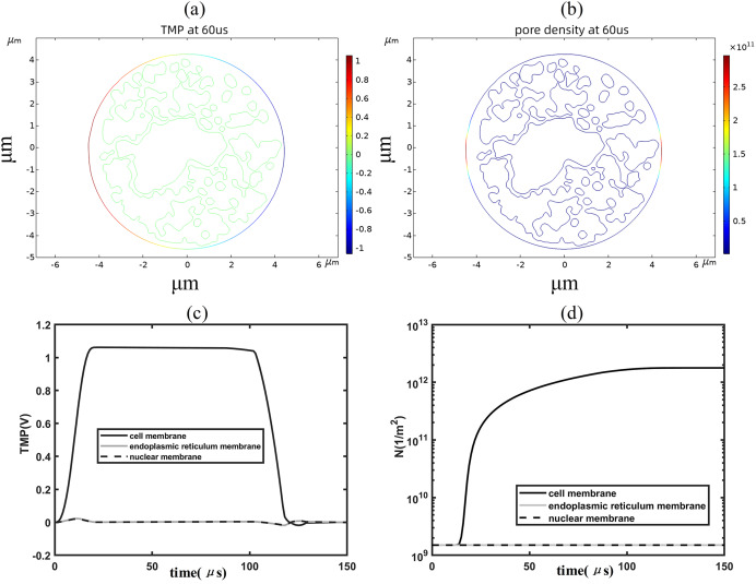 Figure 11
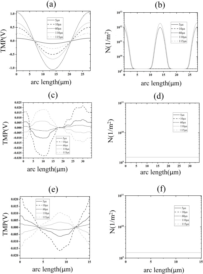 Figure 12
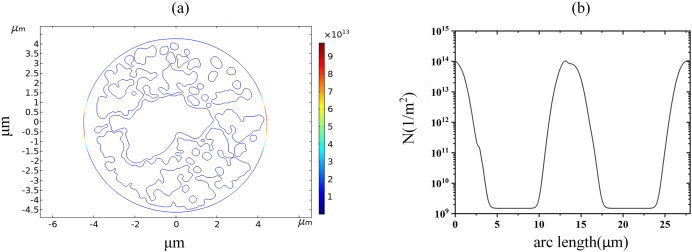 Figure 13
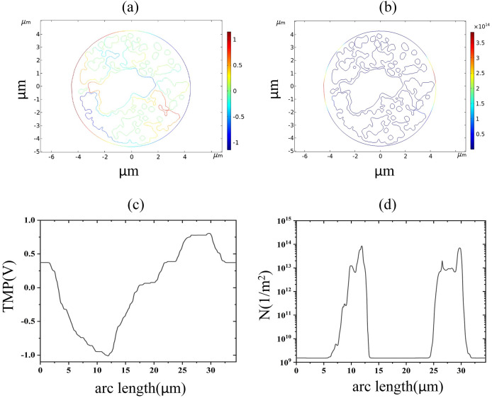 Figure 14
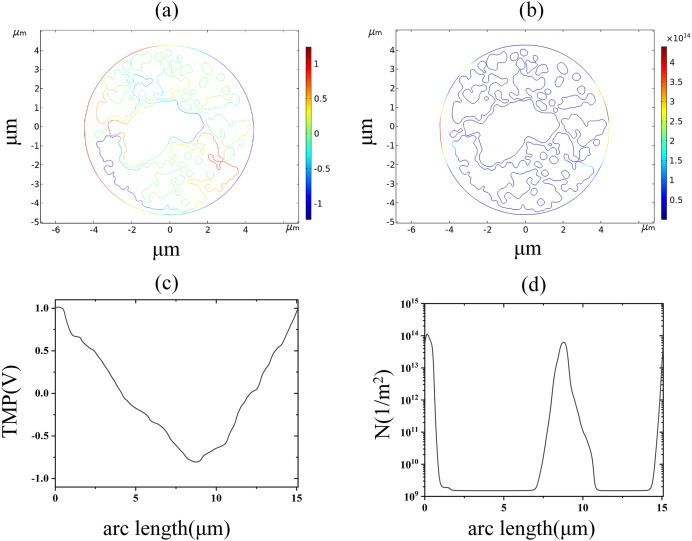 Figure 15
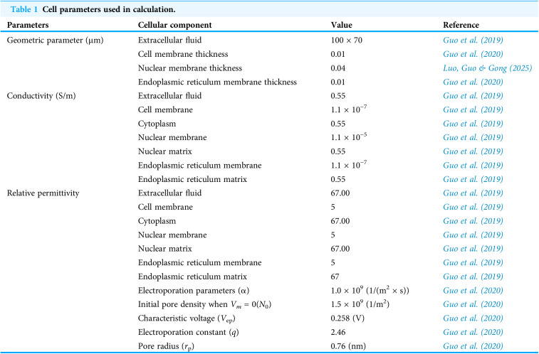 Figure 16
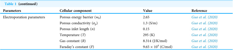 Figure 17
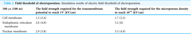 Figure 18
 Figure 19
 Figure 20
Peer Reviews
No public reviews on file for this paper yet. If you reviewed it on a platform where reviews are public (OpenReview, ICLR, NeurIPS, ICML), you can paste yours below so the community can read it here.
Videos
No videos yet. Explain this paper in a talk, walkthrough, or lecture? Add one.
Taxonomy
TopicsMicrobial Inactivation Methods · Microfluidic and Bio-sensing Technologies · Plasma Applications and Diagnostics