High-resolution diffusion imaging in the unfixed post-mortem infant brain at 7 T
Wenchuan Wu, Sebastian W. Rieger, Luke Baxter, Eleri Adams, Jesper L.R. Andersson, Maria M. Cobo, Foteini Andritsou, Matteo Bastiani, Ria Evans Fry, Robert Frost, Sean Fitzgibbon, Sean Foxley, Darren Fowler, Chris Gallagher, Amy F.D. Howard, Joseph V. Hajnal, Fiona Moultrie

TL;DR
This study shows that high-resolution diffusion MRI can be used on unfixed infant brains after death to study brain development.
Contribution
The paper introduces a novel approach for scanning unfixed post-mortem infant brains at ultra-high magnetic field strength.
Findings
High-quality diffusion MRI data was successfully acquired from unfixed post-mortem infant brain tissue.
Altered diffusion properties were observed consistent with post-mortem changes.
Preliminary analyses showed comparable results to in vivo data from age-matched subjects.
Abstract
Diffusion MRI of the infant brain allows investigation of the organizational structure of maturing fibers during brain development. Post-mortem imaging has the potential to achieve high resolution by using long scan times, enabling precise assessment of small structures. Technical development for post-mortem diffusion MRI has primarily focused on scanning of fixed tissue, which is robust to effects like temperature drift that can cause unfixed tissue to degrade. The ability to scan unfixed tissue in the intact body would enable post-mortem studies without organ donation, but poses new technical challenges. This paper describes our approach to scan setup, protocol optimization, and tissue protection in the context of the Developing Human Connectome Project (dHCP) of neonates. A major consideration was the need to preserve the integrity of unfixed tissue during scanning in light of energy…
Genes, proteins, chemicals, diseases, species, mutations and cell lines named across the full text — each resolved to its canonical identifier and authoritative record.
Click any figure to enlarge with its caption.
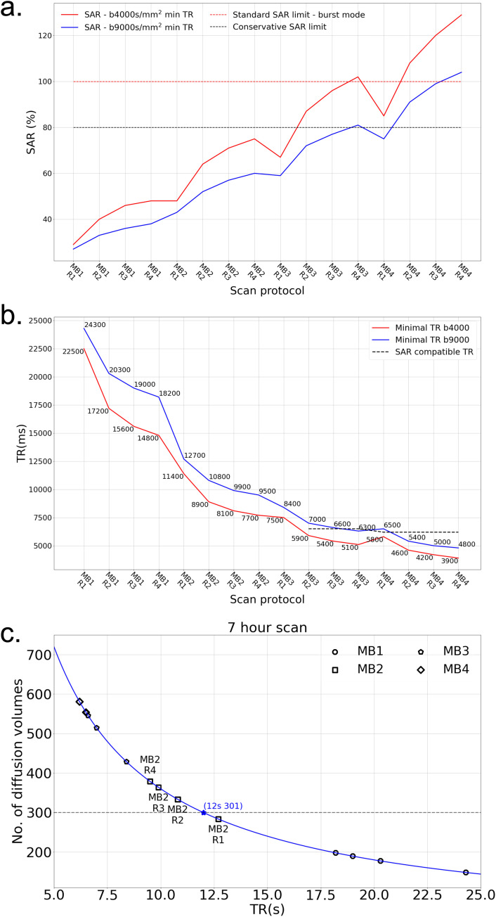 Figure 1
 Figure 2
 Figure 3
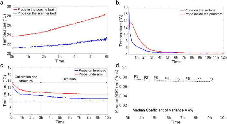 Figure 4
 Figure 5
 Figure 6
 Figure 7
 Figure 8
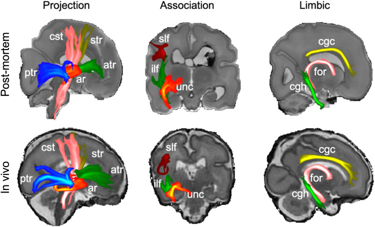 Figure 9
Peer Reviews
No public reviews on file for this paper yet. If you reviewed it on a platform where reviews are public (OpenReview, ICLR, NeurIPS, ICML), you can paste yours below so the community can read it here.
Videos
No videos yet. Explain this paper in a talk, walkthrough, or lecture? Add one.
Taxonomy
TopicsAdvanced Neuroimaging Techniques and Applications · Fetal and Pediatric Neurological Disorders · Advanced MRI Techniques and Applications