Spontaneous expulsion of a huge appendiceal fecalith after endoscopic treatment
Fan Wang, Hongling Wang, Sikai Chen, Qiu Zhao, Jiali Hu

Abstract
Genes, proteins, chemicals, diseases, species, mutations and cell lines named across the full text — each resolved to its canonical identifier and authoritative record.
Click any figure to enlarge with its caption.
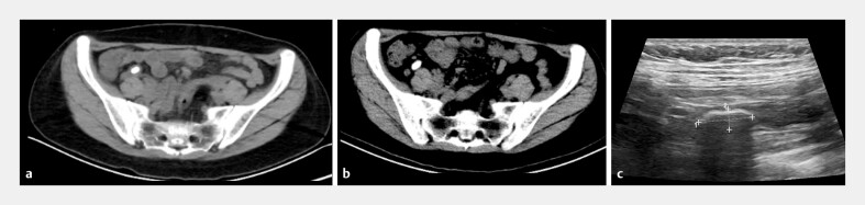 Fig. 1
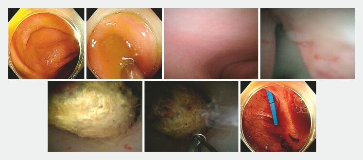 Fig. 2
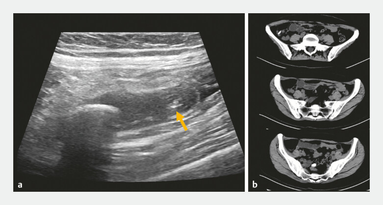 Fig. 3
Peer Reviews
No public reviews on file for this paper yet. If you reviewed it on a platform where reviews are public (OpenReview, ICLR, NeurIPS, ICML), you can paste yours below so the community can read it here.
Videos
No videos yet. Explain this paper in a talk, walkthrough, or lecture? Add one.
Taxonomy
TopicsGastrointestinal disorders and treatments · Diverticular Disease and Complications · Esophageal and GI Pathology
A 38-year-old woman was admitted due to intermittent right lower abdominal pain experienced for half a year. At the local hospital, abdominal computed tomography (CT) scan showed a huge appendiceal fecalith (1.35 × 0.81 cm) ( Fig. 1 ). After admission, abdominal ultrasonography confirmed the appendiceal fecalith (1.49 × 0.58 cm; 3.3 cm from the orifice; appendix size 6.7 × 0.7 cm, retroileal location; diameter 0.8 cm; wall thickness 0.12 cm). Endoscopic retrograde appendicitis therapy using an appendoscope (eyeMAX, 9-French; Micro-Tech [Nanjing] Co., Ltd., China) was planned 1 .
A huge appendiceal fecalith was detected on imaging. a Computed tomography (CT) scan half a year before endoscopic treatment. b Repeat CT scan before endoscopic treatment. c Ultrasonography before endoscopic treatment.
During the operation, the appendoscope was inserted into the appendiceal lumen, and the mucosa was smooth ( Fig. 2 , Video 1 ). Lumen stenosis was detected, and a guidewire was used for exploration before dilating the stenosis repeatedly with the appendoscope body. The fecalith was found at the end of the appendix but could not be grasped with a basket (diameter 1.0 cm). Finally, a plastic stent (8.5 Fr × 5 cm) was implanted into the appendix from the ileocecum.
Endoscopic treatment of the appendiceal fecalith. The appendoscope was passed through the appendiceal orifice and stenosis with the help of a guidewire. The fecalith was detected but could not be grasped with a basket. A plastic stent was placed.
Spontaneous expulsion of a huge appendiceal fecalith after endoscopic treatment.Video 1
Right lower abdominal pain was noted during the following 3 days. On the 4th day, the patient’s pain was significantly relieved, and simultaneous CT scan showed expulsion of the appendiceal fecalith into the sigmoid colon ( Fig. 3 ). On the 11th day, the stent was expelled with the stool. To the best of our knowledge, this is the first reported spontaneous expulsion of a huge appendiceal fecalith after endoscopic treatment.
Imaging after endoscopic treatment. a Ultrasonography detected the fecalith (1.57 × 0.64 cm) and stent (arrow) 2 days after endoscopic treatment. b Computed tomography scan detected the stent end around the appendiceal orifice and fecalith expulsion into the sigmoid colon (size 1.46 × 0.86 cm) 4 days after endoscopic treatment.
Endoscopy_UCTN_Code_TTT_1AQ_2AF
The reference list from the paper itself. Each links out to its DOI / PubMed record.