Endoscopic ultrasonography-guided vascular intervention for bile duct–jejunal anastomotic variceal bleeding
Takashi Kondo, Kazuo Hara, Nozomi Okuno, Shin Haba, Takamichi Kuwahara, Minako Urata, Yoshitaro Yamamoto

Abstract
Genes, proteins, chemicals, diseases, species, mutations and cell lines named across the full text — each resolved to its canonical identifier and authoritative record.
Click any figure to enlarge with its caption.
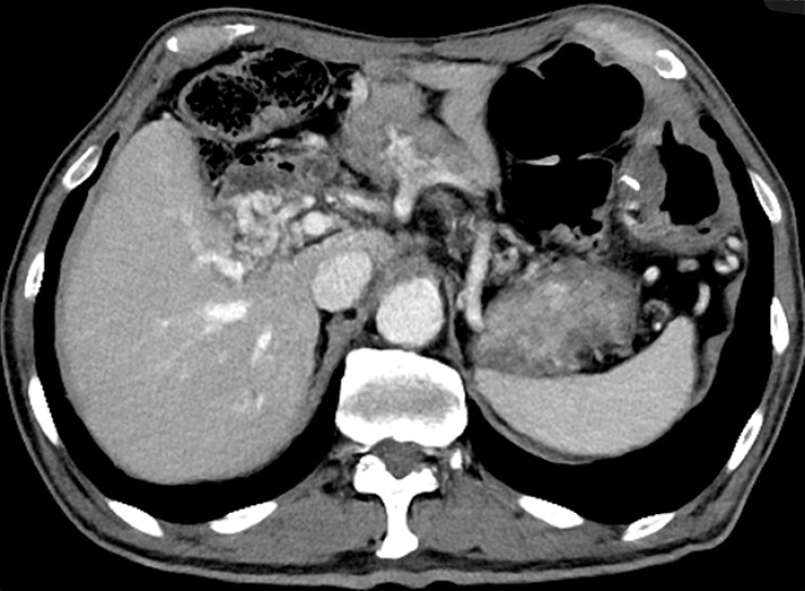 Fig. 1
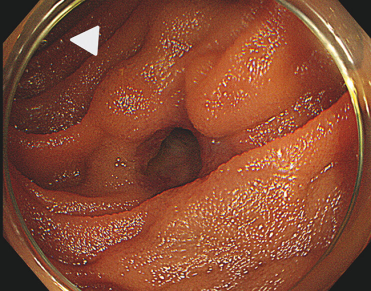 Fig. 2
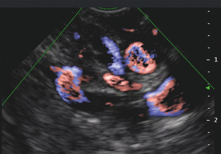 Fig. 3
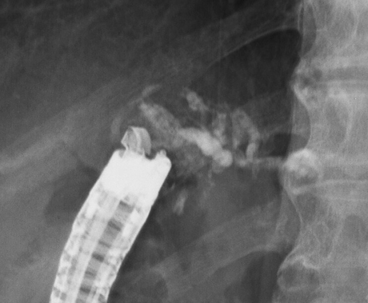 Fig. 4
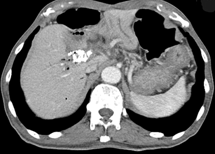 Fig. 5
Peer Reviews
No public reviews on file for this paper yet. If you reviewed it on a platform where reviews are public (OpenReview, ICLR, NeurIPS, ICML), you can paste yours below so the community can read it here.
Videos
No videos yet. Explain this paper in a talk, walkthrough, or lecture? Add one.
Taxonomy
TopicsLiver Disease and Transplantation · Liver Disease Diagnosis and Treatment · Organ Transplantation Techniques and Outcomes
Extrahepatic portal venous obstruction rarely causes ectopic varices in the duodenum or small intestine. Ectopic varices formed by extrahepatic portal venous obstruction can cause bleeding, which is difficult to treat 1 , but there have been several case reports of successful hemostasis using endoscopic ultrasonography (EUS)-guided vascular intervention 2 3 4 . In this study, we report a case in which bile duct–jejunal anastomotic variceal bleeding was successfully stopped using an EUS-guided vascular intervention.
The patient was a 79-year-old man who had undergone pancreaticoduodenectomy and combined portal vein resection for pancreatic cancer. He presented with dark red stool, and blood sampling revealed progressive anemia. A computed tomography (CT) scan showed worsening stenosis of the superior mesenteric vein due to tumor growth and the development of collateral blood vessels around the bile duct–jejunal anastomosis ( Fig. 1 ). He was therefore thought to be experiencing a variceal hemorrhage, owing to tumor-induced extrahepatic portal venous obstruction caused by postoperative recurrence.
Abdominal computed tomography image showing worsening stenosis of the superior mesenteric vein due to tumor growth and the development of collateral blood vessels around the bile duct–jejunal anastomosis.
When an endoscope (CF H290I; Olympus, Tokyo, Japan) was inserted, a bulge of varices was observed around the bile duct–jejunal anastomosis ( Fig. 2 ). Because we were able to observe them using the endoscope, we chose to perform an EUS-guided vascular intervention. The endoscope was removed and a forward-viewing echoendoscope (TGF-UC260J; Olympus Tokyo, Japan) was reinserted. The forward-viewing echoendoscope, which can be easily advanced into the surgically altered small bowel 5 , enabled us to visualize the varices around the anastomosis ( Fig. 3 ; Video 1 ). We punctured the varices with a 19G needle (EZshot3; Olympus, Tokyo, Japan), and injected a mixture of 1.5 mL Histoacryl and 0.5 mL Lipiodol ( Fig. 4 ). A contrast-enhanced CT scan 3 months after the treatment showed the Histoacryl embolizing the varicose vein ( Fig. 5 ). No complications occurred during treatment. No rebleeding was observed prior to the patient’s death 6 months later from pancreatic cancer.
Endoscopic image showing a bulge around the bile duct–jejunal anastomosis (white arrowhead).
Endoscopic ultrasonography image showing the varicose veins around the anastomosis.
Endoscopic ultrasonography-guided vascular intervention is performed to treat bile duct–jejunal anastomotic variceal bleeding.Video 1
Fluoroscopic image of the injected mixture of Histoacryl and Lipiodol.
Contrast-enhanced computed tomography scan 3 months after the treatment showing the Histoacryl embolization of the varicose vein.
EUS-guided vascular intervention can be considered an effective treatment for bile duct–jejunal anastomotic varices.
Endoscopy_UCTN_Code_TTT_1AS_2AH
The reference list from the paper itself. Each links out to its DOI / PubMed record.
- 1Lebrec D Benhamou JP Ectopic varices in portal hypertension Clin Gastroenterol 1985141051213872747 · pubmed ↗
- 2Okuno N Hara K Haba S Endoscopic ultrasound-guided vascular intervention for pancreaticojejunal variceal bleeding Endoscopy 20245632933010.1055/a-2291-9619 PMC 1100381538594005 · doi ↗ · pubmed ↗
- 3Mann R Goyal H Perisetti A Endoscopic ultrasound-guided vascular interventions: Current insights and emerging techniques World J Gastroenterol 2021276874688710.3748/wjg.v 27.i 40.687434790012 PMC 8567466 · doi ↗ · pubmed ↗
- 4Levy I Binmoeller KFEUS-guided vascular interventions Endosc Ultrasound 2018722823510.4103/eus.eus_32_1830117484 PMC 6106145 · doi ↗ · pubmed ↗
- 5Bakr A Hara K Elshair M Utility of forward-view endoscopic ultrasound in fine-needle aspiration in patients with a surgically altered upper gastrointestinal anatomy Clin Endosc 20235636737436600657 10.5946/ce.2021.238PMC 10244154 · doi ↗ · pubmed ↗