Detection and Determinants of Leptospira Infection in Rodents, Cattle, and Humans in Muheza District, Tanzania: A Call for One Health Approach
Gamba Gerald Manyama, Gerald Dickson Mlowe, Athumani Msalale Lupindu, Abdul Suleman Katakweba

TL;DR
This study found Leptospira infections in rodents, cattle, and humans in Tanzania, highlighting the need for a One Health approach to control the disease.
Contribution
The study identifies co-circulating Leptospira serovars and risk factors in a One Health context in Muheza, Tanzania.
Findings
Leptospira seroprevalence was 6.0% in rodents, 12.5% in cattle, and 13.1% in humans.
Farmers were more likely to be infected with Leptospira compared to other occupations.
Serovars Hebdomadis, Sokoine, and Grippotyphosa were most common across species.
Abstract
Interaction among humans, livestock, and wildlife plays an important role in zoonotic disease transmission. The emergence of Leptospira in humans, rodents, and cattle remains relatively understudied. A cross‐sectional study was conducted between February and May 2023 in Muheza to determine evidence of Leptospira infection and associated factors in rodents, cattle, and humans. A total of 479 serum samples from rodents (n = 201), humans (n = 198), and cattle (n = 80) were examined by microscopic agglutination test (MAT) to detect antibodies against 6 live Leptospira stock culture serovars, including Pomona, Hebdomadis, Canicola, Grippotyphosa, Sokoine, and Lora. Additionally, a questionnaire survey was conducted on 140 respondents to determine factors that are associated with Leptospira seropositivity. Descriptive statistics and Chi‐square test were used to analyze the data. The overall…
Genes, proteins, chemicals, diseases, species, mutations and cell lines named across the full text — each resolved to its canonical identifier and authoritative record.
Click any figure to enlarge with its caption.
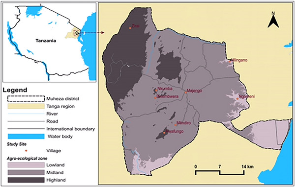 FIGURE 1
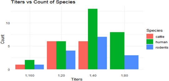 FIGURE 2
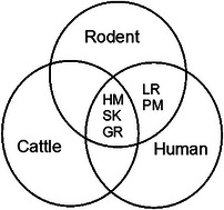 FIGURE 3
| Species | No. of captured and tested | Sex | No of positive | % of positive within species | |
|---|---|---|---|---|---|
| F | M | ||||
|
| 7 | 2 | 5 | 1 | 0.5 |
|
| 2 | 0 | 2 | 0 | 0.0 |
|
| 51 | 19 | 32 | 3 | 1.5 |
|
| 126 | 67 | 59 | 8 | 4.0 |
|
| 15 | 8 | 7 | 0 | 0.0 |
| Total | 201 | 96 | 105 | 12 | 6.0 |
| Parameters | Categories | Number positives | Percentage prevalence |
|---|---|---|---|
| Sex | Cow | 3 | 3.8 |
| Bull | 7 | 8.8 | |
| Grazing pattern | Extensive | 7 | 8.8 |
| Zero | 3 | 3.8 | |
| Location (division) | Amani | 1 | 1.3 |
| Bwembwera | 1 | 1.3 | |
| Muheza | 3 | 3.8 | |
| Ngomeni | 5 | 6.3 |
| Variable | Category | No. and (%) of participants examined | No. and % of participants tested seropositive | Chi‐square ( |
|
|---|---|---|---|---|---|
| Sex | Female | 96 (48.5) | 12 (6.06) | 0.656 | 0.418 |
| Male | 102 (51.5) | 14 (7.07) | |||
| Occupation | Business | 46 (23.2) | 10 (5.05) | 4.771 | 0.549 |
| Employee | 26 (13.1) | 0 (0.00) | |||
| Farmers | 100 (50.5) | 15 (7.58) | |||
| Students | 26 (13.1) | 1 (0.50) | |||
| Location | Amani | 33 (16.7) | 4 (2.02) | 2.115 | 0.549 |
| Bwembwera | 17 (8.6) | 3 (1.50) | |||
| Muheza | 60 (30.3) | 7 (3.50) | |||
| Ngomeni | 88 (44.4) | 12 (6.06) | |||
| Age | 18–35 | 107 (54.0) | 10 (5.05) | 48.864 | 0.284 |
| 36–59 | 80 (40.4) | 15 (7.58) | |||
| ≥60 | 11 (5.6) | 1 (0.50) |
| Species | Rodents | Humans | Cattle | |||
|---|---|---|---|---|---|---|
| Serovars |
| % |
| % |
| % |
| Pomona | 1 | 0.50 | 1 | 0.51 | 0 | 0.00 |
| Hebdomadis | 2 | 1.00 | 7 | 3.54 | 4 | 5.00 |
| Canicola | 0 | 0.00 | 0 | 0.00 | 0 | 0.00 |
| Grippotyphosa | 4 | 1.99 | 12 | 6.06 | 5 | 6.25 |
| Sokoine | 7 | 3.48 | 9 | 4.55 | 4 | 5.00 |
| Lora | 1 | 0.50 | 1 | 0.51 | 0 | 0.00 |
| Total | 15 | 7.47 | 30 | 15.17 | 13 | 16.25 |
- —African Centre of Excellence for Innovative Rodent Pest Management and Biosensor Technology Development (ACE II IRPM‐BTD)
Peer Reviews
No public reviews on file for this paper yet. If you reviewed it on a platform where reviews are public (OpenReview, ICLR, NeurIPS, ICML), you can paste yours below so the community can read it here.
Videos
No videos yet. Explain this paper in a talk, walkthrough, or lecture? Add one.
Taxonomy
TopicsLeptospirosis research and findings
Introduction
1
Leptospirosis is a neglected tropical disease (NTD) of public health importance caused by the pathogenic spirochete that belongs to the genus Leptospira [1]. Leptospira pathogens occur in a variety of forms known as serovars, which are differentiated based on their antigenic characteristics [2]. Globally, approximately 250 Leptospira pathogenic serovars have been identified. The disease has been identified as a global public health problem in animals and humans worldwide [3, 4]. It is estimated that severe human cases between 300,000 and 500,000 occur annually, with a mortality rate of up to 30% [5]. Approximately 58,900 human deaths are reported annually [5]. The disease contributes significantly to morbidity and mortality in many sub‐Saharan African countries and disproportionately affects the poor living in rural communities [3]. According to Allan et al. [1], acute human leptospirosis has been recorded in 18 African nations, including Tanzania.
Rodents are among reservoir hosts of Leptospira worldwide [6]. Domestic animals, such as cattle, are considered the most important reservoir of Leptospira, thus an essential source for human transmission [7]. The common route of transmission to humans is direct or indirect contact with urine or other non‐salivary body fluids of infected domesticated pets and reservoir animals like dogs, pigs, horses, racoons, and wild animals, or contact with contaminated water, soil, or food of infected animals. [8, 9].
In humans, the clinical presentation of the disease is biphasic, with the initial phase being septicemic or acute, and is characterized by a febrile illness lasting almost a week. In the immune phase, antibodies are produced, and leptospires are excreted in the urine [10]. Additionally, the disease can result in miscarriage, stillbirth, maternal death, intrauterine fetal death (IUFD), and congenital infection [11]. In sub‐Saharan countries, about one‐third of pregnant women are estimated to be infected with an NTD agent [12, 13, 14]. Similarly, children under 5 years of age, adults older than65, people with compromised immunity, those with HIV and co‐infections, occupational risks (abattoir workers, veterinarians, farmers, soldiers, and meat vendors), and cancer patients under chemotherapy are at higher risk of Leptospira infection [15]. Leptospirosis can be a very severe disease in humans, with a case fatality rate of 5%–15% if left untreated [5].
Tanzania is regarded as one of many countries with favorable environments for survival of Leptospira because of its tropical climate [7]. Around 10 serovars have been documented in Tanzania. Of these, six—Sokoine (Icterohaemorrhegia), Serjoe, Hebdomadis, Grippotyphosa, Ballum, and Australis—have been detected across humans, livestock, and wild ungulates in the Katavi and Rukwa regions [16]. Other serovars, including Lora, Canicola, and Pomonanhave, been isolated from cattle and rodents in Morogoro areas [17]. The annual incidence of human leptospirosis is estimated to be 75–102 cases per 100,000 population [18]. About 70% of Tanzanians are engaged in livestock keeping, farming, and fishing activities; thus, they are at high risk of Leptospira infection [19]. In recent years, sub‐Saharan African countries, including Tanzania, have experienced periodic outbreaks of human and animal leptospirosis, the most recent being human leptospirosis (Mgunda in Swahili) in Ruangwa, Lindi, whereby three deaths and twenty confirmed cases were reported [7]. Other regions in Tanzania, such as Katavi, have reported a 29.96% prevalence of human leptospirosis [16]. Similarly, 8.8% prevalence in Moshi [20], 13.0% in Kilosa district [21], 12.16% in Mwanza [14], 7.9% in Ngorongoro Conservation Area [22], and 15.1% in Tanga [23]. Additionally, a 13.0%–30.3% prevalence of bovine leptospirosis has been reported in Tanga [24, 25]. Moreover, the prevalence of Leptospira antibodies in rodents ranges from 1.8% to 25.8% in Tanzania [26]. The previous research in the study area focused solely on Leptospira infection in cattle [23, 24, 27], whereas the present study expands the research spectrum to include humans and rodents. The current study has used six available circulating serovars: Sokoine (Icterohaemorrhegia), Hebdomadis, Grippotyphosa, Canicola, Lora, and Pomona. The result of this study provides crucial information on important components of Leptospira transmission cycle and suggests options in design of leptospirosis control plans, including multisectoral, One Health approach.
Materials and Methods
2
Description of the Study Area
2.1
The study was conducted in Muheza district, Tanga (Figure 1). Muheza is one of eight districts of the Tanga region located in north‐eastern Tanzania, at latitudes 40 54′ 18″ S and longitudes 38 0.55′23″ E. The district has a human population of 238,260 and an annual population growth rate of 3.6%, according to the census of 2022 by United Republic of Tanzania [28]. Muheza district is divided into four administrative divisions: Ngomeni, Muheza, Amani, and Bwembwera, and it has a total of 37 wards.
Map of Muheza districts, Tanga region, Tanzania, showing the study sites. Source: QGIS. 3.34.1‐Prizren; EPSG: 4326‐WGS 84.
Agriculture remains the mainstay of the district's economy, with over 79% of households involved in agricultural activities. These households depend on rainfed maize, oranges, rice, and vegetables as their main crops, but livestock keeping is another major economic activity. The cattle population in Muheza district is 27,861, of which 9613 are dairy cattle and the rest are indigenous (Muheza District Council).
The study sites were selected on the basis of the agroecological zone, categorized as lowland (75 m and below), midland (75–300 m), and highland (300 m and above). In these agroecological zones, four divisions, including Bwembwera (Kwafungo, Bwembwera, and Nkumba), Muheza (Majengo, Masuguru, Kwemkabala, and Mtindiro), Ngomeni (Mlingano, Mkuzi, Mkanyageni, and Ngomeni), and Amani (Amani, Zirai), were selected (Figure 1). The hospitals were selected on the basis of their proximity to different agroecological zones within the study locality. Eight villages in each division were purposely chosen based on the agroecological zones and the presence of various habitat types (cropland, peridomestic, and indoor). This selection was made to ensure a more comprehensive representation of the study area's overall community of rodents, cattle, and human settlements.
Study Design and Sample Size Estimation
2.2
This study employed a cross‐sectional design conducted after the rainy season, from February to May 2023, which involved rodents, cattle, and human sampling. The sample size was estimated using the equation developed by (Cochran WG; [29]). n=Z2PQd2 [30]. Whereby n is the sample size, Z = 1.96 (desired confidence level was 95%), P is the proportional factor (15.5% for rodents, 15.1% for humans, and 5.6% for cattle) (Schoonman and Swai [25]), Q is the 1 − p, and d is the desired level of precision (5%). The target was to sample 25 rodents from each of the 8 villages, 10 cattle from each village, and 198 human subjects from Teule Hospital and Ubwari Health Centre. A total of 201 rodents, 198 humans, and 80 cattle were enrolled in this study. One hundred and forty respondents were involved in a face‐to‐face questionnaire interview; among them, 40 were livestock keepers, whereas the rest were patients with the age of 18 and above who visited Teule Hospital and Ubwari Health Centre.
Inclusion and Exclusion Criteria
2.3
The study included patients of all ages with febrile illness who visited Teule Hospital and Ubwari Health Centre, live rodents, and cattle older than 1 year. Humans unwilling to consent were excluded from the study.
Rodent Trapping
2.4
From March 7 to April 17, 2023, rodent trapping was carried out in houses and around human dwellings, farms proximal to human settlements such as cultivated cropland by using Sherman live traps (7.5 × 9.0 × 23.0 cm^3^) and locally made live traps with wooden box wire mesh window (12 × 15 × 20 cm^3^) [31]. A total of 250 traps (200 Sherman live traps and 50 local traps) were placed per site in 10 lines each with 10 trapping stations, 10 m apart in each station and each line for four consecutive nights. Traps were daily baited by using a mixture of peanut butter and maize brans. In cropland, traps were positioned near holes, irrigation canals, and rat trails. In indoor trapping, traps were placed in feed areas, kitchens, and shelves where food was stored [32]. The traps were set at 1700 h evening and inspected early in the morning at 07:00 h. Captured rodents were collected, anesthetized with diethyl ether preserved with ethanol, and identified to species level using the established taxonomic nomenclature. Rodent morphometric data, including weight, total length, tail length, hind foot length, and ear length, were then recorded [13, 33].
Blood Sampling From Rodents and Cattle
2.5
From rodent capture, 1–2 mL of blood was aseptically collected from the heart puncture or plexus by using sterile syringes and needles.
In cattle, which were randomly sampled from different herds from March 17 to 30, 2023, 4 to 10 mL of blood was aseptically collected from the jugular using sterile syringes and needles after proper manual animal restraint.
The samples were immediately transferred into plain vacutainer tubes and allowed to clot for serum separation at room temperature for at least 30 min [34]. The samples were centrifuged for 10 min at 3000 rpm to increase the serum volume. The harvested sera were subsequently transferred into well‐labeled Eppendorf tubes and then stored at −20°C until subjected to microscopic agglutination test (MAT) [33].
Study Humans and Blood Sample Collection
2.6
The present study was carried out among individuals with febrile illness who were visiting Teule Hospital and Ubwari Health Center. The participants were asked for consent to take part in the study and that their blood would be screened for leptospirosis. A total of 198 blood samples were collected from consented subjects who visited Teule Hospital and Ubwari Health Centre from May 15 to 26, 2023, for various diagnostic tests, including malaria and typhoid. A 2–4 mL blood sample was aseptically collected from each study subject by medical personnel using sterile syringes [33]. Socio‐demographic information of study participants, including sex, occupation, location, and age, was recorded.
Microscopic Agglutination Test
2.7
Leptospira stock cultures of the serovars Pomona, Hebdomadis, Canicola, Grippotyphosa, Sokoine, and Lora were purified by sub‐culturing into Ellinghausen–McCullough–Johnson–Harris (EMJH) medium. Pure Leptospira cultures were sub‐cultured and incubated for 5–7 days at 30°C before conducting MAT. The purity of the Leptospira serovars was checked by dark field microscope for adequate density and the absence of contaminating bacteria. The recommended maximum Leptospira density for MAT is 3 × 10^8^ cells/mL on the MacFarland scale [33]. All wells of a microtiter plate were filled with 50 µL phosphate‐buffered saline (PBS), pH 7.2. Except the wells of row 2 that contained 90 µL of PBS. Ten microliters of sera samples were added to the wells of row 2 (dilution was 1:10), followed by serial double dilution with PBS to acquire initial dilutions of 1:10, 1:20, 1:40, 1:80, and 1:160 by pipetting 50 µL from the wells of row 2 to the next rows. At last, the remaining 50 µL were discarded. Afterward, volumes of 50 µL of Leptospira antigen were added to all wells of the microtiter plate for initial screening [35]. All plates were incubated for 2 h at 30°C before reading the results. The antigen–serum mixtures were observed under a dark field microscope by taking a drop of antigen PBS mixture to a microscopic slide. Positive samples titer was recorded by detecting 50% Leptospira agglutination and living 50% of cells free [36]. Additionally, the sample was considered positive for the infection if it had MAT titers of 1:160 in humans. Similarly, samples with titers of 1:20 and 1:80 were considered exposed to Leptospira bacteria. In contrast, samples with MAT titers greater than 1:40 in cattle were regarded as positive. Missed agglutinations were recorded as negative.
Statistical Analysis
2.8
Descriptive statistics, such as proportions and percentages for different variables, were computed in Excel. Seroprevalence was determined as a proportion of number of positive samples to the number of samples tested. Chi‐square test was used to assess the association of seropositivity to different variables such as sex, occupation, residence/location, and age category in humans and species in rodents. A p value of less than 0.05 was regarded as significant.
Ethical Approval
2.9
The research ethical clearance for conducting this study was approved by the Research Ethics Committee at Sokoine University of Agriculture (Ref. No. SUA/ADM/R.1/8/967) issued on December 29, 2022. The permission to conduct research in Muheza was obtained from the Medical Research Coordinating Committee of the National Institute for Medical Research with reference number NIMR/HQ/R.8a/Vol. IX/4269 issued on April 18, 2023. Moreover, before commencing data collection in the study area, authorization was granted from all local authorities, including Muheza district's medical officer (DMO) and village chairmen. Human participants were verbally informed that their blood might be collected and tested for leptospirosis, and verbal consent was obtained from them.
Results
3
Rodent Species Captured in the Study Area
3.1
A total of 201 rodents were captured from indoor, farmland, and peridomestic habitats. Five different species of rodents, including Acomys spp., Lemniscomys spp., Mastomys natalensis, Rattus rattus, and Tatera spp., were captured. Out of the rodents sampled, 96 were females. Remarkably, R. rattus was the most frequent species captured in the study area. On the contrary, the least relative abundances were found in Lemniscomys spp.
Seroprevalence of Leptospira Antibodies in Rodent Species
3.2
The overall seroprevalence of Leptospira antibodies in this study was 6.0% (12/201; 95% CI: 3.12%–10.20%). The highest infection rate was found in R. rattus at 4.0% (8/201), followed by M. natalensis at 1.5% (3/201), and the lowest was in Acomys spp. as shown in Table 1. However, the difference in seropositivity between rodent species was not statistically significant (p = 0.3318). Further, the study revealed the variation of seroprevalence of Leptospira serovars among rodents to habitat types (p < 0.00) and locations (*p *< 0.000) (Table 1).
Seroprevalence of Leptospira Antibodies in Cattle in the Study Area
3.3
The seroprevalence of Leptospira antibodies in cattle was 12.5% (10/80; 95% CI: 6.16%–21.79%). The variation of seropositivity for sex, grazing system, and location is shown in Table 2.
Seroprevalence and Socio‐Demographic Characteristics of Humans
3.4
The seroprevalence and socio‐demographic characteristics of humans are shown in Table 3. It was found that out of 198 participants, 48.5% (96/198) were females. The rest were males. The seroprevalence was 13.1% (95% CI: 8.76%–18.65%). A seroprevalence of 7.58% (15/198) recorded among the farmers was significantly high compared to other occupations (χ ^2 ^= 9.1894, df = 3, p = 0.02688).
Seroprevalence of Different Leptospiral Serovars in Rodents, Humans, and Cattle
3.5
Analysis of leptospiral serovars shows that the predominant serovar in humans was Grippotyphosa at 6.06% (12/198), whereas serovar Canicola was not detected (Table 4). In rodents, the predominant serovar was Sokoine at 3.48% (7/201), and serovar Canicola was not detected. Similarly, the predominant serovar in cattle was Grippotyphosa at 6.25% (5/80), whereas serovars Pomona, Canicola, and Lora were not detected (Table 4). Furthermore, the statistical analysis shows that there was no statistically significant difference in the seroprevalence of Leptospira serovars among cattle to grazing pattern (χ ^2 ^= 1.847, df = 1, p = 0.1741), age (χ ^2^ = 4.4976, df = 9, p = 0.8757), location (χ ^2 ^= 5.0286, df = 3, p = 0.1697), and sex (χ ^2 ^= 1.6162, df = 1, p = 0.2036). A higher seropositive proportion was observed in female cattle than in males (Table 2). Similarly, a higher percentage of seropositive cases was observed in extensive grazing areas (R) compared to the zero‐grazing pattern (Z) (Table 2).
Proportions of Leptospira Antibody Titers in Cattle, Humans, and Rodents
3.6
The highest antibody level observed was 1:160. This level was found in two human samples for the Sokoine serovar and one sample each for the Grippotyphosa and Pomona serovars in rodents and cattle. However, the 1:160 level was not found in the Hebdomadis, Canicola, and Lora serovars. The most common antibody level was 1:40, which was most frequently observed in humans. The 1:60, 1:40, and 1:80 were also commonly found in humans, whereas the 1:20 level was most common in both humans and cattle. The 1:80 level was not found in cattle (Figure 2). Serovar Grippotyphosa showed high frequencies for all titers 4.59% (n = 22), followed by serovar Sokoine 4.18% (n = 20) (Table 4). Serovar Canicola was not detected in the study area.
Proportion of Leptospira antibody titers in cattle, humans, and rodents in Muheza.
Multiple Reactions of Leptospira Serovars in Rodents, Humans, and Cattle
3.7
Out of 479 rodents, cattle, and humans’ sera that were tested against 6 Leptospira serovars, 8 (1.67%) serum samples reacted with more than 1 serovar. In this study, multiple reactions in rodents, cattle, and humans occurred with serovars Hebdomadis, Sokoine, and Grippotyphosa. Furthermore, multiple reactions between rodents and humans occurred with serovars Lora and Pomona only, and none was observed in cattle. No multiple reactions with serovar Canicola were found in any of the three hosts (Figure 3).
Multiple reactions of Leptospira serovars in rodents, humans, and cattle.
Comparison of Seroprevalence of Leptospiral Infection Among Different Variables
3.8
Overall, the statistical analysis showed that the variation of seroprevalence of Leptospira serovars among rodents, humans, and cattle was significant (χ ^2 ^= 6.0046, df = 2, p = 0.04967). However, individual species did not show significant differences: rodents (χ ^2 ^= 2.3713e − 30, df = 1, p = 1), cattle (χ ^2^ = 2.7517, df = 1, p = 0.09715), and humans (χ ^2 ^= 0.001994, df = 1, p = 0.9644).
Discussion
4
The present study has found that the seroprevalence of Leptospira antibodies in rodents is 6.0%. This finding is higher than 1.9% reported earlier [37] and lower than 20.29% reported from Katavi region [16] and 16.9% in Morogoro [38]. On the other hand, the seroprevalence of 12.5% in cattle in the present study is lower than 30.37% reported in Katavi [16] and higher than 6.6% reported in Kigoma and Kagera regions [40]. The seropositivity of 13.1% in humans in the current study is lower than 29.96% from Katavi [16], 30.1% in Endulen, Tanzania [22], and 29.9% in Katavi [39]. On the other hand, the results of the present study are higher than 10.75% from a study in Morogoro [38] and 11.1% reported from Kigoma and Kagera regions [40]. In all these reports, MAT was the method used to determine the seroprevalence. The differences in seropositivity in rodents, cattle, and humans could be due to variation in ecology of the study sites. Moreover, there were some differences in Leptospira reference serovars used, which might have led to the differences in the outcome. The current study in Muheza, Tanga, used six references serovars: Sokoine, Lora, Pomona, Hebdomadis, Canicola, and Grippotyphosa. A study in Morogoro [38] also used six serovars: Sokoine, Kenya, Mwogolo, Lora, Canicola, and Grippotyphosa. In a study in Kigoma and Kagera, seven serovars were used, which include Sokoine, Kenya, Sejroe, Grippotyphosa, Bataviae, Lora, and Pomona. In another study in Morogoro, Kilimanjaro, Mbeya, Tanga, Singida, Mwanza, Ruvuma, and Rukwa [37], six Leptospira serovars, namely, Sokoine, Harjo, Canicola, Pyogenes, icterohaemorrhagiae and Grippotyphosa, were used. The use of one serovar in one study and not in another could be a possible cause of the differences.
The current study has involved rodents (wildlife), cattle (livestock), and humans. Antibodies against Hebdomadis, Grippotyphosa, and Sokoine serovars were detected in all host species, that is, rodents, cattle, and humans. This study differs from previous studies in study area [23, 27], which investigated seroprevalence of Leptospira antibodies in cattle only. As a result, the burden, risk population, and transmission dynamics were difficult to quantify. The current study is similar to studies conducted on rodents, livestock, humans, and wildlife in Katavi–Rukwa ecosystem [16], Bahi, Dodoma [13], and Kilombero, Morogoro [40]. This scenario gives a clue that there is a possibility of sharing of Leptospira exposure, something that was previously speculated [13]. Therefore, the findings of the present study call for One Health approach in planning and implementation of Leptospira control, whereby personnel from veterinary, medical, wildlife, and environmental sectors and the community have to team up.
The present study has reported multiple agglutinations in 8 sera samples (1.67%) in rodent, cattle, and humans by MAT. MAT is a reference diagnostic tool for Leptospira infection. It can discriminate current or active infection from chronic exposure, but it cannot distinguish between co‐infection and cross‐reaction in the case of multiple reactions [41, 42, 43]. In the present study, the serum with highest antibody titer in multiple coagulations was assumed to be the infecting serovar, similar to procedures in other studies [44]. In other words, it was not possible to tell if the multiple coagulations represented multiple infections or an infection and cross‐reaction. To resolve this misnomer, the isolation of Leptospira by culture method or molecular typing is recommended.
Detection of Leptospira antibodies in M. natalensis and Acomys spp. in this study underscores the risk to public health. Similar findings have been reported in Kibondo and Kakonko, Kigoma [45]. These rodent species live outside the human settlement and can easily come into contact with livestock or contaminate the environment. As such, they may be a potential source of human leptospirosis [1, 38]. Therefore, there is a need for control of rodents within and around human residence.
The current study found that farmers had the higher Leptospira seroprevalence than other occupations, indicating a higher likelihood of being infected with Leptospira than other groups. This is especially so, considering the close human contact with animals (rodents and cattle), when sharing common environment; watering points, habitats, and unsecured feeds [46]. The finding of this study is in‐line with other previous studies that have highlighted farmers as being the highest risk groups for leptospirosis in parts of Tanzania and elsewhere [22, 23, 47]. Other researchers have highlighted various groups, including dog keepers [48], livestock keepers [24], fishing communities [33], abattoir workers [49, 50], as well as miners and sewage workers [47], being the other risk groups for contracting leptospirosis Swai, E.S., Schoonman, L., & Daborn, C., [51]. Therefore, there is a need for creation of awareness on this occupational risk to farmers.
The present study has revealed that the predominant serovar stimulating antibody production in humans was Grippotyphosa, followed by Sokoine. This finding is in agreement with reports from studies on predominance of these serovars in humans [13, 14, 23, 24, 47]. Grippotyphosa serovar is naturally habored by cattle [50], whereas rodents are well‐known reservoirs of Sokoine serovar [13]. The detection of antibodies for Grippotyphosa and Sokoine serovars in humans as well as in cattle and rodents suggests the presence of close contact among humans, cattle, and rodents, and possibly sharing of the bacteria. Therefore, in order to reduce this public health threat, rodent control strategies and education to cattle keepers on Leptospira infection risk are recommended.
Different levels of antibody titers have been reported in this study. The 1:40 titer was most abundant in humans compared to other titers. This suggests that humans have been exposed to a specific serovar, triggering the production of IgM at high concentration. A lower titer in humans implies a chronic exposure to Leptospira that is associated with the presence of IgM [52]. On the other hand, the higher titer of 1:160 was observed in two sera of humans, one rodent, and one cattle serum. This suggests that there was active or acute Leptospira infection [38]. The presence of both chronic and acute exposures in the study area implies that Leptospira infection risk is a long‐standing threat that calls for intervention.
Limitations of the Study
4.1
In Africa, the highest numbers of panel serovars in detecting Leptospira antibodies by MAT is 10 serovars [14]. The current study may have underestimated the seroprevalence of Leptospira antibodies because only six serovars were included in the panel.
Conclusion and Recommendations
4.2
Cattle, rodents, and humans in the Muheza district are infected with five out of the six circulating Leptospira serovars, including possible incidences of co‐infection. The findings give a clue on the complexity of a possible Leptospira transmission cycle in the study area and other similar areas.
The detection of high Leptospira antibodies in rodents, cattle, and humans suggests acute infection. We recommend a multisectoral One Health approach in control of Leptospira infection that involves veterinary, medical, wildlife, and environmental personnel as well as the community through rodent control measures, public awareness campaigns, occupational safety protocols for farmers, and other workers who are at great risk of exposure and infection.
Author Contributions
Gamba Gerald Manyama: writing – original draft, conceptualization, methodology, data curation, software, investigation, validation, formal analysis, project administration, funding acquisition, writing – review and editing, visualization, resources. Gerald Dickson Mlowe: software, data curation, validation, formal analysis. Athumani Msalale Lupindu: conceptualization, methodology, data curation, investigation, validation, supervision, visualization, writing – review and editing, project administration, software. Abdul Suleman Katakweba: conceptualization, methodology, investigation, validation, formal analysis, supervision, project administration, resources, funding acquisition, visualization, writing – review and editing.
Conflicts of Interest
The authors declare no conflicts of interest.
The reference list from the paper itself. Each links out to its DOI / PubMed record.
- 1K. J. Allan , H. M. Biggs , J. E. B. Halliday , and R. R. Kazwala , “Epidemiology of Leptospirosis in Africa: A Systematic Review of a Neglected Zoonosis and a Paradigm for ‘One Health’ in Africa,” PLOS Neglected Tropical Diseases 9, no. 9 (2015): e 0003899, 10.1371/journal.pntd.0003899.26368568 PMC 4569256 · doi ↗ · pubmed ↗
- 2S. N. Ahmad , S. Shah , and F. M. H. Ahmad , “Laboratory Diagnosis of Leptospirosis,” Journal of Postgraduate Medicine 51 (2005): 195–200.16333192 · pubmed ↗
- 3P. R. Torgerson , J. E. Hagan , F. Costa , et al., “Global Burden of Leptospirosis: Estimated in Terms of Disability Adjusted Life Years,” PLOS Neglected Tropical Diseases 9, no. 10 (2015): e 0004122, 10.1371/journal.pntd.0004122.26431366 PMC 4591975 · doi ↗ · pubmed ↗
- 4P. Vijayachari , A. P. Sugunan , and A. N. Shriram , “Leptospirosis: An Emerging Global Public Health Problem,” Journal of Biosciences 33, no. 4 (2008): 557–569, 10.1007/s 12038-008-0074-z.19208981 · doi ↗ · pubmed ↗
- 5F. Costa , J. E. Hagan , J. Calcagno , et al., “Global Morbidity and Mortality of Leptospirosis: A Systematic Review,” PLOS Neglected Tropical Diseases 9, no. 9 (2015): e 0003898, 10.1371/journal.pntd.0003898.26379143 PMC 4574773 · doi ↗ · pubmed ↗
- 6K. Boey , K. Shiokawa , and S. Rajeev , “ Leptospira Infection in Rats: A Literature Review of Global Prevalence and Distribution,” P Lo S Neglected Tropical Diseases 13, no. 8 (2019): 1–24, 10.1371/journal.pntd.0007499.PMC 668878831398190 · doi ↗ · pubmed ↗
- 7D. S. Masunga , A. Rai , M. Abbass , et al., “Leptospirosis Outbreak in Tanzania: An Alarming Situation,” Annals of Medicine and Surgery 80 (2022): 104347, 10.1016/j.amsu.2022.104347.35992205 PMC 9382409 · doi ↗ · pubmed ↗
- 8V. Barragan , S. Olivas , P. Keim , and T. Pearson , “Critical Knowledge Gaps in Our Understanding of Environmental Cycling,” Applied and Environmental Microbiology 83, no. 19 (2017): 1–10.10.1128/AEM.01190-17PMC 560134628754706 · doi ↗ · pubmed ↗