Correction: S-propargyl-cysteine attenuates temporomandibular joint osteoarthritis by regulating macrophage polarization via Inhibition of JAK/STAT signaling
Wenyi Cai, Antong Wu, Zhongxiao Lin, Wei Cao, Janak L. Pathak, Richard T. Jaspers, Rui Li, Xin Li, Kaihan Zheng, Yufu Lin, Na Zhou, Xin Zhang, Yizhun Zhu, Qingbin Zhang

Abstract
Genes, proteins, chemicals, diseases, species, mutations and cell lines named across the full text — each resolved to its canonical identifier and authoritative record.
Click any figure to enlarge with its caption.
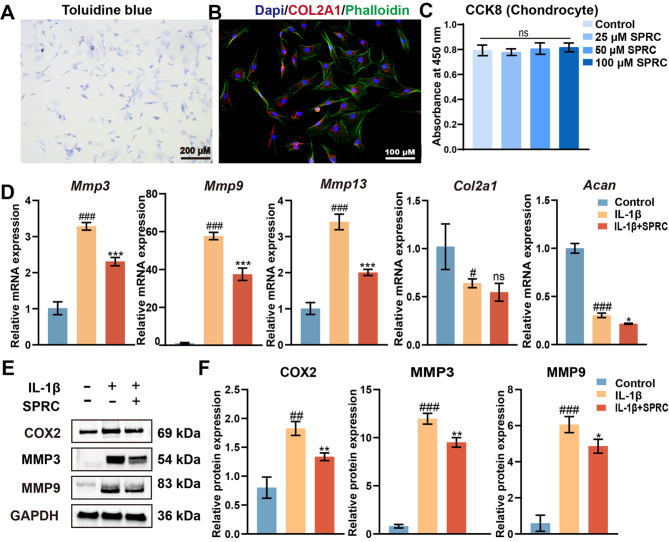 Figure 1
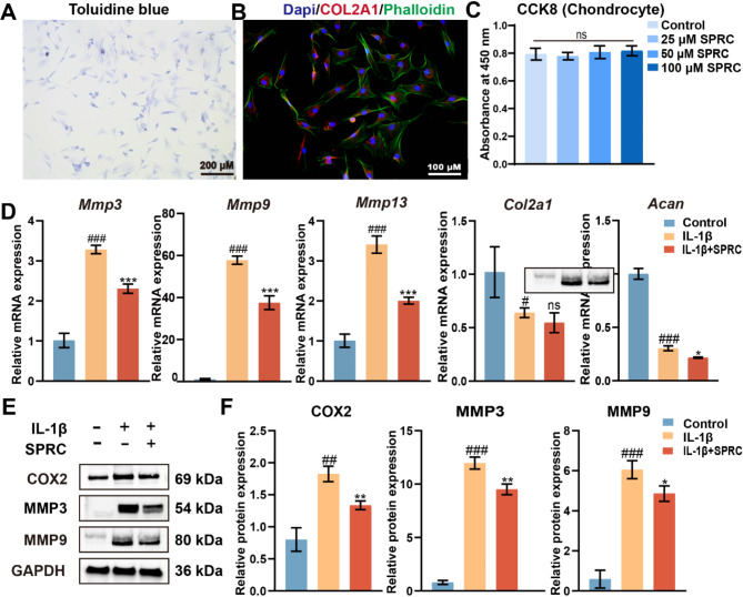 Figure 2
Peer Reviews
No public reviews on file for this paper yet. If you reviewed it on a platform where reviews are public (OpenReview, ICLR, NeurIPS, ICML), you can paste yours below so the community can read it here.
Videos
No videos yet. Explain this paper in a talk, walkthrough, or lecture? Add one.
Taxonomy
TopicsOsteoarthritis Treatment and Mechanisms · Cytokine Signaling Pathways and Interactions · Mast cells and histamine
Correction: Molecular Medicine (2025) 31:128
10.1186/s10020-025-01186-6
In this article, Fig. 6 appeared incorrectly and have now been corrected in the original publication. For completeness and transparency, both correct and incorrect versions are displayed below.
Incorrect Fig. 6.
Fig. 6SPRC reduced condylar chondrocytes ECM catabolism in vitro. (A) Representative images of condylar chondrocytes stained with toluidine blue to assess extracellular matrix (ECM) integrity. (B) Immunofluorescence staining of chondrocytes showing DAPI-stained nuclei (blue), phalloidin-stained F-actin (green), and COL2A1 (red). (C) Cell viability of rat primary condylar chondrocytes (rPCCs) treated with SPRC (25, 50, or 100 μM) for 24 h, as evaluated by the CCK8 assay. (D) RT-qPCR analysis of matrix degradation-related genes (Mmp3, Mmp9, and Mmp13) and matrix synthesis-related genes (Col2a1 and Acan). (E-F) Western blot analysis of COX2, MMP3, and MMP9 protein expression levels. The data were analyzed via one-way ANOVA (n ≥ 3). Statistical significance is denoted as follows: #p<0.05, ##p < 0.01, and ###p < 0.001 compared with the control group; *p < 0.05, **p < 0.01, and ***p < 0.001 compared with the IL-1β group. “ns” indicates no significance
Correct Fig. 6.
Fig. 6SPRC reduced condylar chondrocytes ECM catabolism in vitro. (A) Representative images of condylar chondrocytes stained with toluidine blue to assess extracellular matrix (ECM) integrity. (B) Immunofluorescence staining of chondrocytes showing DAPI-stained nuclei (blue), phalloidin-stained F-actin (green), and COL2A1 (red). (C) Cell viability of rat primary condylar chondrocytes (rPCCs) treated with SPRC (25, 50, or 100 μM) for 24 h, as evaluated by the CCK8 assay. (D) RT-qPCR analysis of matrix degradation-related genes (Mmp3, Mmp9, and Mmp13) and matrix synthesis-related genes (Col2a1 and Acan). (E-F) Western blot analysis of COX2, MMP3, and MMP9 protein expression levels. The data were analyzed via one-way ANOVA (n ≥ 3). Statistical significance is denoted as follows: #p<0.05, ##p < 0.01, and ###p < 0.001 compared with the control group; *p < 0.05, **p < 0.01, and ***p < 0.001 compared with the IL-1β group. “ns” indicates no significance
The original article has been corrected.