Swallow Syncope as a Late Complication of Sleeve Gastrectomy
Fred Rudensky, Sana N Khan, Prasad Chalasani

TL;DR
A rare case of swallow syncope is reported as a late complication of sleeve gastrectomy, linked to a hiatal hernia and successfully treated with a pacemaker.
Contribution
This is the second reported case of swallow syncope following sleeve gastrectomy and highlights the role of iatrogenic hiatal hernia as a potential cause.
Findings
Swallow syncope occurred six weeks after sleeve gastrectomy and was associated with a progressively worsening hiatal hernia.
The patient's symptoms resolved after dual-chamber pacemaker placement despite unattempted lifestyle or medical interventions.
The case suggests a possible hereditary component and raises questions about post-infectious changes contributing to the condition.
Abstract
Swallow syncope, also known as swallow-induced syncope, or deglutition syncope, is a type of situational reflex syncope associated with swallowing. It is believed to be due to exaggerated vagal parasympathetic stimulation leading to inhibition of heart rate during swallowing. Swallow syncope has been documented in cases of gastroesophageal structural pathologies, such as achalasia and esophageal stricture, and has even been shown to resolve following surgical correction. To date, there are 118 reported cases of swallow syncope published in medical literature, which includes 14 reports of swallow syncope with associated hiatal hernia, and only one of which reports swallow syncope following sleeve gastrectomy. We present a case of swallow syncope as a late complication of laparoscopic sleeve gastrectomy associated with gradually worsening hiatal hernia. Our patient is a 54-year-old…
Genes, proteins, chemicals, diseases, species, mutations and cell lines named across the full text — each resolved to its canonical identifier and authoritative record.
Click any figure to enlarge with its caption.
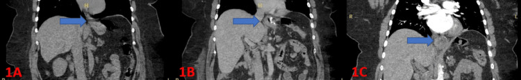 Figure 1
Peer Reviews
No public reviews on file for this paper yet. If you reviewed it on a platform where reviews are public (OpenReview, ICLR, NeurIPS, ICML), you can paste yours below so the community can read it here.
Videos
No videos yet. Explain this paper in a talk, walkthrough, or lecture? Add one.
Taxonomy
TopicsCardiovascular Syncope and Autonomic Disorders · Obstructive Sleep Apnea Research · Sympathectomy and Hyperhidrosis Treatments
Introduction
Swallow syncope, also known as swallow-induced syncope, or deglutition syncope, is a type of situational reflex syncope associated with swallowing [1]. There have been a total of 117 cases of swallow syncope reported in the literature [2]. It is believed to be due to exaggerated parasympathetic vagal nerve signaling to the sinoatrial and atrioventricular nodes, leading to the suppression of heart rate during deglutition [2,3]. Lower esophageal mechanoreceptors are stimulated by the passing of food or liquid bolus causing afferent signaling to the adjacent motor nucleus, leading to efferent vagal nerve parasympathetic signaling causing suppression of heart rate and contractility [3]. Swallow syncope has been documented in cases of gastroesophageal structural pathologies, such as achalasia and esophageal stricture, and has even been shown to resolve following surgical correction of the underlying gastroesophageal issues [2]. To date, there are at least 19 reports of swallow syncope with associated hiatal hernia [2,4], and at least one report of swallow syncope following vertical sleeve gastrectomy [5]. We present a case of swallow syncope as a late complication of laparoscopic sleeve gastrectomy with subsequent gradually increasing hiatal hernia confirmed on CT imaging.
Case presentation
Our patient is a 54-year-old female who presented with a chief complaint of episodic presyncope and syncope that occurs when swallowing food or liquids. Pertinent past medical history included laparoscopic sleeve gastrectomy complicated by surgical infection six weeks prior to presentation, morbid obesity, obstructive sleep apnea, and a remote history of neurocardiogenic syncope during childhood. The patient described the childhood neurocardiogenic syncope as one or two episodes of syncope due to emotional distress which did not require intervention, but could not elaborate further. The patient described her presenting symptoms as sudden fogginess, dizziness, and loss of consciousness lasting approximately 30-45 seconds. She stated the episodes were not caused by any specific foods and began approximately 10 seconds after swallowing. She also stated that her smart watch alerted her to bradycardia on multiple occasions but could not recall the lowest recorded rate. She denied any other symptoms associated with the syncopal episodes including chest pain, palpitations, abdominal pain, or diaphoresis. She denied tobacco, alcohol, or drug use. Of note, the patient reported her twin sister had undergone Roux-en-Y gastric bypass procedure and subsequently developed swallow syncope. Swallow syncope was formally diagnosed, although it is not known if she had any anatomical structural abnormalities such as hiatal hernia.
Review of home prescriptions did not show any medications which increased risk of bradyarrhythmia. Prior medical records revealed a recent transthoracic echocardiogram showing normal ventricular function and an ejection fraction of approximately 60% without evidence of wall motion abnormalities or structural defects. Electrocardiogram from date of admission showed sinus bradycardia with a heart rate of 55 beats per minute with no other abnormal findings and no significant changes from prior studies. Doppler ultrasound of the carotid arteries showed minimal plaque formation with patent flow bilaterally. Review of EKGs and vital signs from prior studies did reveal occasional sinus bradycardia with heart rates ranging from 50-60 beats per minute. Although attempts at reproducing patient’s syncopal episodes under visual observation were not made due to safety concerns, nursing staff did report witnessing multiple episodes. Asystolic pauses lasting three to four seconds and sinus bradycardia with heart rates as low as 20 beats per minute were recorded on telemetry monitoring during the patient’s hospital stay. All of the asystolic pauses and episodes of bradycardia below 50 beats per minute occurred exclusively during ingestion of food or liquid. The patient’s respiratory rate and blood pressure remained stable and within normal limits during these episodes.
CT Angiogram of the chest did not reveal any acute cardiopulmonary process, but did show a hiatal hernia adjacent to the left atrium (Figure 1). No further imaging studies were deemed necessary at that time, although later review of CT imaging from the patient’s prior admissions revealed that the hiatal hernia had gradually increased in size since the first appearing on CT imaging from five weeks prior. The hiatal hernia was not present on imaging prior to her bariatric procedure (Figure 1). No pertinent laboratory abnormalities were noted. The patient was found to have markedly low heart rate variability, as well as minimal change to heart rate when transitioning from recumbent position to standing and ambulating, indicating possible autonomic dysfunction although formal testing with tilt table test or deep breathing test was not conducted. Following evaluation by electrophysiology consult, a shared decision was made to proceed with dual chamber pacemaker placement.
CT imaging showing progressive hiatal herniaA: CT of the abdomen and pelvis showing absence of a hiatal hernia (blue arrow) 13 months prior to the patient's laporascopic sleeve gastrectomy.B: CT of the abdomen and pelvis showing presence of a hiatal hernia (blue arrow) eight days after the patient's laparoscopic sleeve gastrectomyC: CT angiogram of the chest showing progressive enlargement of a hiatal hernia (blue arrow) two months after the patient's laparoscopic sleeve gastrectomy
As per the anesthesia report from the date of pacemaker implantation, the patient’s heart rate was 58 immediately prior to administration of midazolam, propofol, and succinylcholine. Following anesthesia induction and intubation, the patient’s sinus bradycardia gradually worsened although rhythm remained sinus. Once the atrial lead was advanced to the chosen area of implantation, the patient’s rhythm became junctional with a rate of 34 beats per minute requiring temporary pacing set to a rate of 60 beats per minute. The pacer was implanted and a minimum rate of 50 beats per minute was set. No complications were noted following the procedure and the patient quickly recovered. She was discharged the following day and instructed to follow up with electrophysiology consult and her primary care provider.
Review of EKG studies from emergency department visits and hospital admissions for unrelated complaints in the months following pacemaker implantation showed alternating sinus bradycardia and normal sinus rhythm, although heart rate did not decrease below 57 beats per minute. The patient reports still experiencing intermittent bradycardia but states she has not experienced any presyncopal or syncopal episodes seven months post pacemaker placement, although swallow-induced bradycardia was not formally assessed after implantation.
Discussion
In a literature review of 101 cases of swallow syncope, atrioventricular blocks were noted in 34.7% of patients while sinus dysfunction was noted in 33.7% [2]. Of the 101 cases, 32.7% of patients presented with associated gastrointestinal comorbidities [2]. Although there is only one report of swallow syncope associated with bariatric procedure prior to the current case [5], sinus bradycardia is strongly associated with bariatric surgery, with one study estimating approximately 15% of patients developing vasovagal syncope following bariatric procedure [6], and one study estimating prevalence of bradyarrythmia following bariatric procedure to be approximately 18% [7].
While not entirely understood, the central cause of swallow syncope is believed to be excessive parasympathetic stimulation [2]. More specifically, the mechanism behind swallow syncope is suspected to be impulses conducted through reflex arcs between sensory fibers of the esophageal branches and cardiac branches of the vagus nerve leading to inappropriate parasympathetic vagal stimulation with subsequent suppression of cardiac heart rate and contractility [2]. Our patient's development of progressive bradycardia with anesthesia induction supports vagal hypersensitivity as an underlying factor in the pathogenesis of her swallow syncope and the presence of a hiatal hernia with gastric fundus herniating into the space located directly between the heart and esophageal and vagal plexus strongly aligns with this theory. However, no mention of a hiatal hernia was made in the aforementioned report of swallow syncope associated with laparoscopic sleeve gastrectomy [5]. An oropharyngeal mechanism responsible for swallow syncope has also been proposed. In one study, investigators used electrophysiological testing to measure the time between initiation of swallowing and onset of bradycardia or asystole, with results showing the delay to be approximately two to three seconds [8]. This relatively short delay in onset points toward the oropharynx as the location from which initial nerve signaling originates, although it is believed that the oropharyngeal and lower esophageal origination points are a continuum of the same esophageal nerve plexus.
Decrease in BMI leading to metabolic feedback loops has also been suggested as the cause of sinus bradycardia following bariatric surgery [6]. Severity of bradyarrhythmia increased as amount of weight loss postoperatively increased [7,8]. One study found that patients who developed sinus bradycardia following bariatric procedure had an average decrease in BMI of 34.4% [7]. Studies did not distinguish swallow syncope from sinus bradycardia or other bradyarrythmias. In the case of our patient, pre-procedure weight of 135.9 kg decreased significantly to 122.7 kg at time of presentation, with weight loss totaling 13.2 kg (9.02%) over approximately six weeks. The pathophysiology responsible for the correlation between weight loss and severity of bradyarrhythmia is suspected to be changes in GLP-1 and leptin levels [9]. However, a recent meta-analysis of 27 trials including a total of 70,299 patients found no increased risk of atrial or ventricular arrythmias with GLP-1 agonist use [10]. A hereditary component is also possible, as evidenced in our case, as well as one other reported case of swallow syncope [11]. A twin study including 51 same-sex twin pairs showed a significant higher concordance in monozygotic twins than in dizygotic twins in syncope associated with typical vasovagal triggers (p=<0.001) [12].
Few treatment guidelines specific to reflex syncope, which includes the situational forms of syncope such as swallow syncope, have been published. The existing recommendations are based on evidence limited to case reports, case series, and small observational studies [1]. According to the 2017 American College of Cardiology (ACC)/American Heart Association (AHA)/Heart Rhythm Society (HRS) guidelines for the evaluation and management of patients with syncope, treatment of reflex syncope is centered on avoidance of triggers, increased salt and water intake, and reduction or removal of diuretics and antihypertensive medications [1]. Reversible underlying etiology should be corrected whenever possible [1]. Although irritation of the esophageal and vagal plexus due to hiatal hernia is suspected to be responsible for our patient's syncopal events, it was not identified as a correctable cause during the patient's hospital admission. Furthermore, laparoscopic fundoplication for the correction of hiatal hernias is reserved for patients with Type II, III, and IV (paraoesophageal) hernias [13]. Staging is based on barium swallow radiography and is necessary to determine treatment course [13] and fundoplication may not have been the recommended approach in our patient's case. The ACC/AHA/HRS guidelines recommend pacemaker placement for the treatment of recurrent vasovagal syncope with asystolic pauses in patients over the age of 40 (Class IIb evidence), but there are no recommendations made pertaining to pacemaker placement in neuroinhibitory reflex syncope [1]. However, the International Study on Syncope of Uncertain Etiology 3 (ISSUE-3), a randomized double-blinded placebo-controlled trial did find a 32% absolute risk reduction of recurrent syncope following dual chamber pacemaker placement in patients with severe neurally mediated asystolic syncope [14]. All forms of reflex syncope were included, excluding carotid sinus syndrome [14]. The 2018 European Society of Cardiology (ESC) Guidelines for the diagnosis and management of syncope reserves treatment of reflex syncope with cardiac pacing for older patients with the dominant cardioinhibition subtype as evidenced by lack of prodrome [15].
Conclusions
Our case of swallow syncope secondary to a bariatric procedure is one of two such cases reported in medical literature. Aspects of our case such as the progressive development of a large hiatal hernia, family history of swallow syncope secondary to Roux-en-Y procedure in the patient's twin sister, and successful treatment with dual chamber pacemaker placement distinguish it from other reported cases of swallow syncope. Genetic predisposition is possibly involved in the development of our patient's symptoms. Although a hereditary component in syncope has been previously studied, review of the literature revealed a lack of studies pertaining to the development of hiatal herniation, indicating there is a need for retrospective studies investigating for any genetic factors in patients with hiatal hernias. The choice to treat our patient's reflex syncope with dual chamber pacemaker placement was based on the need for immediate resolution of symptoms due to the severe impact her symptoms had on quality of life, although timely identification of the hiatal hernia may have influenced the decision to proceed with cardiac pacing. Clinicians should consider structural anatomical abnormalities and gastrointestinal sources of pathogenesis during their evaluation as this may reveal a reversible cause of syncope. This is especially true for patients presenting with bradyarrythmias following bariatric procedures. Further investigation is warranted to help guide screening and diagnosis.
The reference list from the paper itself. Each links out to its DOI / PubMed record.
- 12017 ACC/AHA/HRS guideline for the evaluation and management of patients with syncope: a report of the American College of Cardiology/American Heart Association Task Force on Clinical Practice Guidelines and the Heart Rhythm Society Heart Rhythm Shen WK Sheldon RS Benditt DG 021714201710.1016/j.hrthm.2017.03.00428286247 · doi ↗ · pubmed ↗
- 2Swallow syncope: a case report and review of literature BMC Cardiovasc Disord Siew KS Tan MP Hilmi IN Loch A 1911920193139100010.1186/s 12872-019-1174-4PMC 6686266 · doi ↗ · pubmed ↗
- 3Cases of swallow syncope induced by the activation of mechanorecepters in the lower esophagus Korean J Intern Med Kang KH Cho WH Kim MC Chang HJ Chung JI Won DJ 68712020051590695610.3904/kjim.2005.20.1.68PMC 3891415 · doi ↗ · pubmed ↗
- 4An interesting case of recurrent postprandial cardiogenic syncope caused by type III hiatal hernia Cureus Chaudhary AJ Qureshi MH El Sharu H Prostak J 015202310.7759/cureus.47791 PMC 1067656738021578 · doi ↗ · pubmed ↗
- 5Swallow syncope after laparoscopic vertical sleeve gastrectomy Surg Obes Relat Dis Rezvani M Casillas SG Antanavicius G 089201310.1016/j.soard.2013.03.00223747073 · doi ↗ · pubmed ↗
- 6Prevalence of vasovagal syncope following bariatric surgery Saudi J Anaesth Al Obeed OA Traiki TB Alfahad YF 1611641520213418863510.4103/sja.SJA_922_20PMC 8191276 · doi ↗ · pubmed ↗
- 7Asymptomatic sinus bradycardia following bariatric surgery Am J Cardiol Malik MG Franklin SM Whigham LA Castellanos A Fontaine JM 1049105311320142446207210.1016/j.amjcard.2013.11.069 · doi ↗ · pubmed ↗
- 8Swallow-induced syncope in 5 patients: electrophysiologic evaluation during swallowing Neurol Clin Pract Aydogdu I Hasdemir C Acarer A Alpaydin S Ertekin C 316323720172918553610.1212/CPJ.0000000000000376 PMC 5648200 · doi ↗ · pubmed ↗