A Rare Case of Primary Follicular Lymphoma in the Retroperitoneum
Tijin Mathew, Shree Hariprasad, Teresa Varghese, Lydia George, Benjamin Easow, Parth Adrejiya, Melinda Gevorgian, Greeshma Thomas, Tehmina Zafar

TL;DR
This paper presents a rare case of follicular lymphoma in the retroperitoneum, emphasizing its unusual symptoms and the importance of early diagnosis.
Contribution
The novelty lies in highlighting an atypical presentation of follicular lymphoma with pleural effusion and ascites, not typically associated with B symptoms.
Findings
The patient presented with abdominal swelling and retroperitoneal adenopathy, leading to a diagnosis of grade II follicular lymphoma.
Follicular lymphoma was detected without typical B symptoms, suggesting it should be considered in differential diagnoses for pleural effusion or ascites.
Early diagnosis and treatment of follicular lymphoma can result in favorable clinical outcomes.
Abstract
Primary retroperitoneal non-Hodgkin lymphoma (NHL) is considered sporadic, difficult to diagnose, and has an atypical presentation. NHL typically presents with non-tender peripheral lymphadenopathy in two-thirds of patients. Very rarely, it may present with abdominal swelling and obstructive symptoms of the respiratory and GI tracts. The primary gastrointestinal NHL accounts for 5-20% of all extra-nodal lymphomas. However, there are only a few reported cases of primary retroperitoneal lymphomas. Among them, most were diffuse large B cell lymphomas. This case is of a 54-year-old male patient who presented to the emergency department with abdominal swelling for the past four months. The CT abdomen and pelvis were indicative of massive retroperitoneal and mesenteric adenopathy with concerns for lymphoma with some volume ascites. He underwent an interventional radiology (IR)-guided…
Genes, proteins, chemicals, diseases, species, mutations and cell lines named across the full text — each resolved to its canonical identifier and authoritative record.
Click any figure to enlarge with its caption.
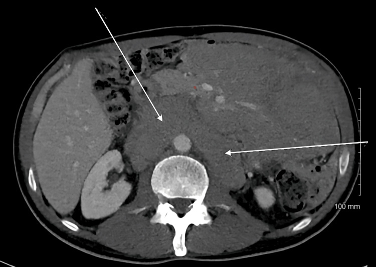 Figure 1
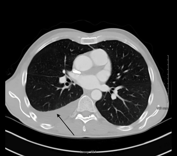 Figure 2
| Component | Reference range | Serum | Pleural fluid | Pleural fluid/Serum |
| Protein (mg/dl) | 6 to 8 | 5.3 | <1.8 | 0.3 |
| LDH (IU/L) | 140 to 271 | 315 | 88 | 0.279 |
| Triglycerides (mg/dl) | <100 | N/A | 2895 | N/A |
| pH of body fluid | 7.35 to 7.45 | N/A | 7.77 | N/A |
| Grades of the follicular lymphomas | Pathology findings |
| Grade 1 | Follicular small cleaved cell lymphoma with 0 to 5 centroblasts per high-power field (HPF) |
| Grade 2 | Follicular mixed–cell lymphoma with 6 to 15 centroblasts per HPF |
| Grade 3 | Follicular large cell lymphoma with >15 centroblasts per HPF |
| Grade 3 A | Centrocytes present |
| Grade 3 B | Solid sheets of centroblasts |
Peer Reviews
No public reviews on file for this paper yet. If you reviewed it on a platform where reviews are public (OpenReview, ICLR, NeurIPS, ICML), you can paste yours below so the community can read it here.
Videos
No videos yet. Explain this paper in a talk, walkthrough, or lecture? Add one.
Taxonomy
TopicsLymphoma Diagnosis and Treatment · Ovarian cancer diagnosis and treatment · Viral-associated cancers and disorders
Introduction
Primary retroperitoneal non-Hodgkin lymphomas (NHL) are considered sporadic and atypical presentations, thus proving to be a diagnostic challenge. Follicular lymphoma is considered to be the second-most common type of NHL, accounting for approximately 20-25% of the cases in the United States and Europe [1,2]. It can present anywhere in the body; however, based on its location, the presenting complaints may vary. Rarely five to 10% of cases may involve the retroperitoneum, often presenting with delayed diagnosis. All the scantily reported retroperitoneal lymphomas were large B-cell lymphomas [2-5]. While NHL typically presents with non-tender peripheral lymphadenopathy, it can occasionally manifest with obstructive symptoms such as pleural effusions or ascites. Here, we report a rare case of follicular lymphoma presenting as primary retroperitoneal NHL, with ascites and pleural effusion as the initial symptoms.
Case presentation
This case involves a 54-year-old male patient with a past medical history significant only for hypertension, who presented to the emergency department with abdominal swelling for the past four months. He reported that he had abdominal distension and slight discomfort in the left upper quadrant. He had some occasional dull abdominal pain involving bilateral lower quadrants with no changes in pain characteristics with food intake or change in position. He had no significant surgical history and denied any unintentional weight loss, fever, and night sweats. He also denied any recent travel or diarrhea as well as any alcohol or smoking history.
The patient's vital signs were stable. The physical examination was unremarkable for any peripheral lymphadenopathy. On examination, he was found to have mild generalized abdominal distension and decreased breath sound to the left lung bases. His complete blood cell count with differential and comprehensible metabolic panel was unremarkable. The CT abdomen and pelvis were concerning for massive retroperitoneal and mesenteric adenopathy with some volume ascites (Figure 1).
CT abdomen showing massive retroperitoneal and mesenteric adenopathy (white arrows)
Given his pleural effusion on the CT abdomen and pelvis, a CT chest was ordered which showed loculated pleural effusion on the left side and trace pleural effusion on the right (Figure 2).
CT chest showed loculated pleural effusion on the left side and trace pleural effusion on the right (black arrow)
He underwent interventional radiology (IR)-guided thoracentesis and 300 cc of opaque white, chylous pleural fluid was drained. The pleural fluid and serum studies are listed in Table 1.
As per the Light's criteria, his etiology was considered as transudative. His differential body fluid showed 100 cells with 16% neutrophils, 53% lymphocytes, and 31% monocytes and macrophages. The pleural fluid culture was unremarkable. The patient also underwent an IR-guided retroperitoneal lymph node biopsy. The biopsy results indicate grade II follicular lymphoma with positivity for CD20, PAX5, BCL2, CD10, and BCL6, and occasional disruption of the CD21 dendritic cell meshwork. The tumor shows no cyclin D1 expression, a Ki67 proliferation index of <10%, and a low perforation rate (<10%). The patient was referred to the Hematology/Oncology department for further management. He completed six cycles of chemotherapy with bendamustine and rituximab. He tolerated the chemotherapy without any complications. On his six-month oncology follow-up, the Positron Emission Tomography (PET) scan showed complete disease resolution.
Discussion
Primary retroperitoneal masses are often asymptomatic and present atypically, with lymphomas accounting for approximately 33% of all retroperitoneal tumors [6]. Among NHLs, diffuse large B-cell lymphoma (DLBCL) is the most common subtype, comprising 25-30% of cases, while follicular lymphoma is the second most prevalent [1,7]. Follicular lymphoma is a slow-progressing malignancy, with a median age of diagnosis of 63 years [8-10].
Chylous ascites and pleural effusion are rare manifestations of lymphoma. The proposed pathophysiological mechanism involves obstruction of the lymphatic drainage due to external compression, leading to leakage from dilated subserosal lymphatic ducts into the pleural and peritoneal cavities. Given its rare location and indolent nature, retroperitoneal follicular lymphoma is often diagnosed late despite its favorable prognosis when detected early.
Contrast-enhanced CT is the imaging modality of choice for diagnosing retroperitoneal lymphoma [8]. Additional imaging techniques include magnetic resonance imaging (MRI) and PET-CT. However, a definitive diagnosis requires tissue sampling. While excisional biopsy is the gold standard, fine needle aspiration (FNA) offers a less invasive and more time-efficient alternative [6]. In this case, FNA of the retroperitoneal mass confirmed the diagnosis of follicular lymphoma.
Follicular lymphomas can be divided into grades based on the pathology findings [9]. This classification is summarized in Table 2.
Treatment options vary based on the stage and the biological behavior of the disease, whether indolent or aggressive. Management strategies include radiotherapy, immunochemotherapy (rituximab plus chemotherapy), the R-CHOP (rituximab, cyclophosphamide, doxorubicin, vincristine, and prednisone) regimen, single-agent rituximab, the CVP (cyclophosphamide, vincristine, and prednisolone) regimen, and combined immunochemotherapy with radiotherapy [10]. In select cases, surgical resection may be considered for localized disease or to alleviate tumor-related symptoms. In Stage I and Grades 1, 2, and 3, radiotherapy alone is preferred [9,10]. With advanced stages and grades, the patient needs aggressive treatment with immunochemotherapy like R-CHOP [9,10].
Conclusions
This case highlights an atypical presentation of primary retroperitoneal follicular lymphoma with chylous ascites and pleural effusion, a rare manifestation that can delay diagnosis. Given its indolent nature, early recognition of follicular lymphomas in unusual locations, even in the absence of B symptoms, is critical for timely intervention. Imaging modalities such as contrast-enhanced CT and PET-CT are integral to the initial evaluation. However, a definitive diagnosis requires histopathological confirmation via biopsy. Treatment is specific to the disease stage and progression, with immunochemotherapy remaining the mainstay of management. This case underscores the importance of including lymphomas in the differential diagnosis of unexplained pleural effusion or ascites and emphasizes the need for prompt oncologic evaluation to optimize patient outcomes.
The reference list from the paper itself. Each links out to its DOI / PubMed record.
- 1Follicular Lymphoma Stat Pearls [Internet] Kaseb H Ali MA Gasalberti DP Koshy NV Treasure Island (FL)Stat Pearls Publishing 2024 https://www.ncbi.nlm.nih.gov/books/NBK 538206/30855794 · pubmed ↗
- 2Obstructive jaundice by a retroperitoneal lymphoma mimics a pancreatic cancer: a case report J Pancreas Jiménez Jiménez María Belvis Belén Maldonado Pérez 151156192018 https://www.primescholars.com/articles/obstructive-jaundice-by-a-retroperitoneal-lymphoma-mimics-apancreatic-cancer-a-case-report.pdf
- 3Diffuse large B-cell lymphoma with primary retroperitoneal presentation: clinico-pathologic study of nine cases Ann Oncol Pileri SA Zinzani PL Ascani S 14451453122001 https://pubmed.ncbi.nlm.nih.gov/11762818/1176281810.1023/a:1012559725243 · doi ↗ · pubmed ↗
- 4Diagnosis of lymphoma by endoscopic ultrasound-assisted transendoscopic direct retroperitoneal lymph node biopsy: A case report (with video)Endosc Ultrasound Guo J Sun B Wang S 697242015 https://pmc.ncbi.nlm.nih.gov/articles/PMC 4362009/2578928910.4103/2303-9027.151368 PMC 4362009 · doi ↗ · pubmed ↗
- 5An uncommon clinical presentation of retroperitoneal non-Hodgkin lymphoma successfully treated with chemotherapy: a case report World J Gastroenterol Fulignati C Pantaleo P Cipriani G Turrini M Nicastro R Mazzanti R Neri B 31513155112005 https://pubmed.ncbi.nlm.nih.gov/15918208/1591820810.3748/wjg.v 11.i 20.3151 PMC 4305858 · doi ↗ · pubmed ↗
- 6Primary retroperitoneal masses: what is the differential diagnosis?Abdom Imaging Scali EP Chandler TM Heffernan EJ Coyle J Harris AC Chang SD 18871903402015 https://pubmed.ncbi.nlm.nih.gov/25468494/2546849410.1007/s 00261-014-0311-x · doi ↗ · pubmed ↗
- 7Diffuse Large B-Cell Lymphoma Stat Pearls [Internet] Padala SA Kallam A Treasure Island (FL)Stat Pearls Publishing 2023 https://www.ncbi.nlm.nih.gov/books/NBK 557796/32491728 · pubmed ↗
- 8CT findings of lymphoma with peritoneal, omental and mesenteric involvement: peritoneal lymphomatosis Eur J Radiol Karaosmanoglu D Karcaaltincaba M Oguz B Akata D Ozmen M Akhan O 313317712009 https://www.sciencedirect.com/science/article/pii/S 0720048 X 080022831851390610.1016/j.ejrad.2008.04.012 · doi ↗ · pubmed ↗