Validation of Quantitative Ultrasound and Texture Derivative Analyses-Based Model for Upfront Prediction of Neoadjuvant Chemotherapy Response in Breast Cancer
Adrian Wai Chan, Lakshmanan Sannachi, Daniel Moore-Palhares, Archya Dasgupta, Sonal Gandhi, Rossanna Pezo, Andrea Eisen, Ellen Warner, Frances C. Wright, Nicole Look Hong, Ali Sadeghi-Naini, Mia Skarpathiotakis, Belinda Curpen, Carrie Betel, Michael C. Kolios, Maureen Trudeau

TL;DR
This study validates a model using ultrasound and texture analysis to predict how breast cancer patients will respond to chemotherapy before treatment begins.
Contribution
The first validation of a model using quantitative ultrasound, texture derivatives, and molecular features to predict neoadjuvant chemotherapy response in breast cancer.
Findings
The model achieved 94% sensitivity and 95% F1 score in predicting chemotherapy response.
Unplanned chemotherapy changes reduced negative predictive value but increased specificity to 100%.
Misclassifications were attributed to chemotherapy modifications, DCIS presence, and unclear tumor borders.
Abstract
This work was conducted in order to validate a pre-treatment quantitative ultrasound (QUS) and texture derivative analyses-based prediction model proposed in our previous study to identify responders and non-responders to neoadjuvant chemotherapy in patients with breast cancer. The validation cohort consisted of 56 breast cancer patients diagnosed between the years 2018 and 2021. Among all patients, 53 were treated with neoadjuvant chemotherapy and three had unplanned changes in their chemotherapy cycles. Radio Frequency (RF) data were collected volumetrically prior to the start of chemotherapy. In addition to tumour region (core), a 5 mm tumour-margin was also chosen for parameters estimation. The prediction model, which was developed previously based on quantitative ultrasound, texture derivative, and tumour molecular subtypes, was used to identify responders and non-responders. The…
Genes, proteins, chemicals, diseases, species, mutations and cell lines named across the full text — each resolved to its canonical identifier and authoritative record.
Click any figure to enlarge with its caption.
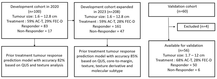 Figure 1
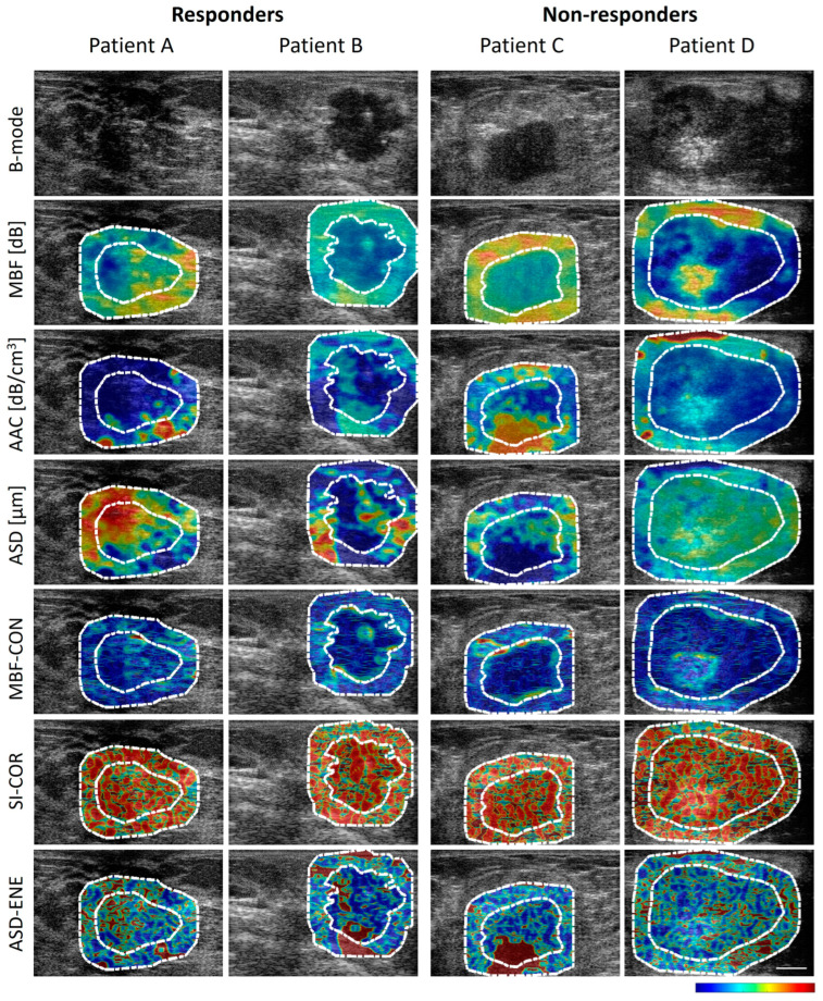 Figure 2
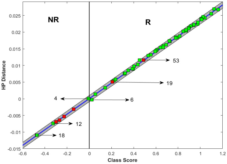 Figure 3
 Figure 4
Peer Reviews
No public reviews on file for this paper yet. If you reviewed it on a platform where reviews are public (OpenReview, ICLR, NeurIPS, ICML), you can paste yours below so the community can read it here.
Videos
No videos yet. Explain this paper in a talk, walkthrough, or lecture? Add one.
Taxonomy
TopicsBreast Cancer Treatment Studies · Breast Lesions and Carcinomas · Radiomics and Machine Learning in Medical Imaging