Correction: Cavazzoni et al. Pemetrexed Enhances Membrane PD-L1 Expression and Potentiates T Cell-Mediated Cytotoxicity by Anti-PD-L1 Antibody Therapy in Non-Small-Cell Lung Cancer. Cancers 2020, 12, 666
Andrea Cavazzoni, Graziana Digiacomo, Roberta Alfieri, Silvia La Monica, Claudia Fumarola, Maricla Galetti, Mara Bonelli, Daniele Cretella, Valeria Barili, Alessandra Zecca, Elisa Giovannetti, Michelangelo Fiorentino, Marcello Tiseo, Pier Giorgio Petronini, Andrea Ardizzoni

Abstract
Genes, proteins, chemicals, diseases, species, mutations and cell lines named across the full text — each resolved to its canonical identifier and authoritative record.
Click any figure to enlarge with its caption.
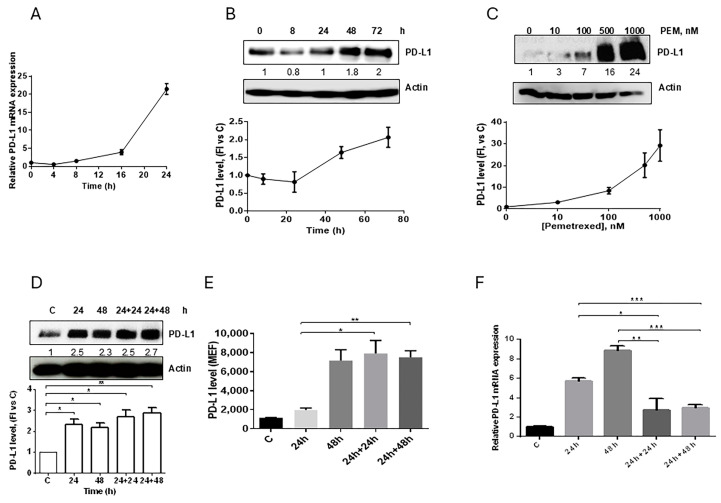 Figure 2
Peer Reviews
No public reviews on file for this paper yet. If you reviewed it on a platform where reviews are public (OpenReview, ICLR, NeurIPS, ICML), you can paste yours below so the community can read it here.
Videos
No videos yet. Explain this paper in a talk, walkthrough, or lecture? Add one.
Taxonomy
TopicsCancer Immunotherapy and Biomarkers · Lung Cancer Research Studies · Cancer Cells and Metastasis
Error in Figure
In the original publication [1], there was a mistake in Figure 2C,D as published. An incorrect image of Actin was inserted as a loading control. The corrected Figure 2 appears below. Additionally, the associated Supplementary Figure S7 has been updated. The authors apologize for any inconvenience caused and state that the scientific conclusions are unaffected. This correction was approved by the Academic Editor. The original publication has also been updated.
Effect of pemetrexed on PD-L1 expression in A549 cell line. (A) A549 cells were treated with 100 nM pemetrexed for the indicated period of time and PD-L1 mRNA level, evaluated by RT-PCR, was reported. (B) Time-dependent modulation (100 nM pemetrexed) and (C) dose-dependent modulation (72 h) of PD-L1 protein expression in A549 cells were evaluated by western blotting. A549 cells were continuously exposed to 500 nM pemetrexed for the indicated period of time or treated for 24 h and, after drug removal, the cells were incubated with fresh medium for 24 h or 48 h. At the indicated times, total PD-L1 protein, membrane PD-L1 protein, and PD-L1 mRNA were quantified by western blotting (D), flow cytometry (E), and RT-PCR (F), respectively. * p < 0.05; ** p < 0.01; *** p < 0.001. Data in (A), (E), and (F) are mean values ± SD of three independent experiments. Results in (B–D) are representative of three independent experiments.
The reference list from the paper itself. Each links out to its DOI / PubMed record.
- 1Cavazzoni A. Digiacomo G. Alfieri R. La Monica S. Fumarola C. Galetti M. Bonelli M. Cretella D. Barili V. Zecca A. Pemetrexed Enhances Membrane PD-L 1 Expression and Potentiates T Cell-Mediated Cytotoxicity by Anti-PD-L 1 Antibody Therapy in Non-Small-Cell Lung Cancer Cancers 20201266610.3390/cancers 1203066632178474 PMC 7139811 · doi ↗ · pubmed ↗