Unconscious Male With Reported Seizure
Naleen Patel, Bradley S. Jackson, Nima Sarani

Abstract
Genes, proteins, chemicals, diseases, species, mutations and cell lines named across the full text — each resolved to its canonical identifier and authoritative record.
Click any figure to enlarge with its caption.
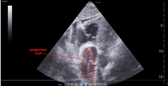 Figure 1
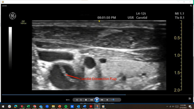 Figure 2
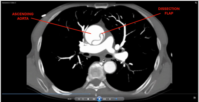 Figure 3
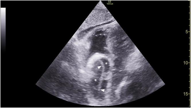 Figure 4
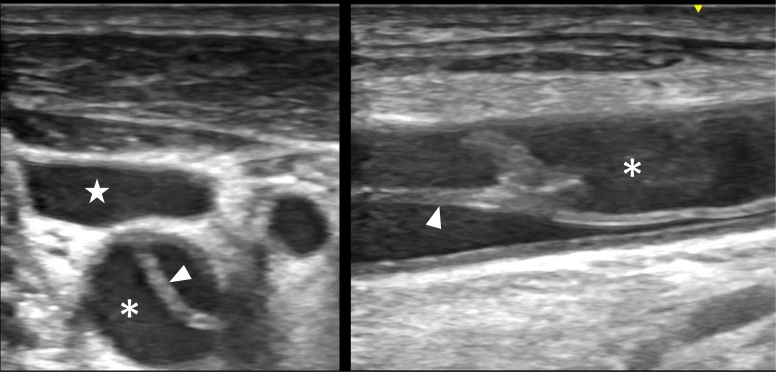 Figure 5
 Figure 6
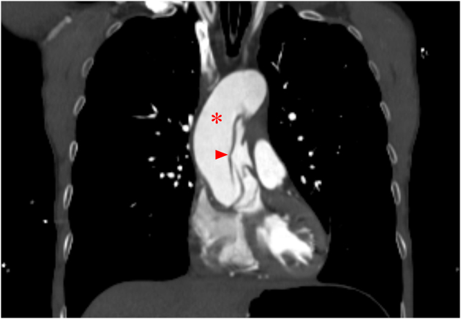 Figure 7
Peer Reviews
No public reviews on file for this paper yet. If you reviewed it on a platform where reviews are public (OpenReview, ICLR, NeurIPS, ICML), you can paste yours below so the community can read it here.
Videos
No videos yet. Explain this paper in a talk, walkthrough, or lecture? Add one.
Taxonomy
TopicsAortic Disease and Treatment Approaches · Traumatic Brain Injury and Neurovascular Disturbances · Cardiac Valve Diseases and Treatments
Case Presentation
1
An unidentified 68-year-old male with an unknown medical history was brought to the emergency department via ambulance after bystanders witnessed the patient suffering seizure-like activity at the library. Intramuscular midazolam was administered prior to arrival. He presented with agonal breathing requiring bag ventilation with a Glasgow Coma Scale score of 3. He was intubated for airway protection. Prior to transport for head imaging, he had a transient episode of postintubation hypotension that prompted a cardiac point-of-care ultrasound examination (Fig 1, Video 1). These findings prompted bedside carotid ultrasound (Fig 2, Video 2).Figure 1. Subxiphoid point-of-care ultrasound showing ascending aorta (asterisks) with an echogenic linear dissection flap (arrowheads).Video 1Subxiphoid point-of-care ultrasound showing ascending aorta with a dissection flap.Figure 2. Transverse-axis (left panel) and long-axis (right panel) views of point-of-care right neck ultrasound demonstrating an internal jugular vein (star), carotid artery (asterisk), and echogenic linear carotid dissection flap (arrowhead).Video 2Transverse-axis point-of-care ultrasound of the right neck demonstrating carotid dissection.
Diagnosis: Stanford Type A Dissection With Bilateral Carotid Extension
2
The subxiphoid view noted a prominent aortic root with direct visualization of a dissection flap. This prompted esmolol initiation and computed tomography angiography imaging of the torso, head, and neck. Stanford type A dissection was confirmed with extension into all branches of the aortic arch, most critically into the left carotid artery with M1 involvement (Figs 3 and 4, Video 3).Figure 3. Transverse computed tomography angiography of the neck showing dissection flaps (arrowheads) in the brachiocephalic (red), left common carotid (yellow), and left subclavian (blue) arteries.Figure 4. Coronal computed tomography angiography of the chest showing an ascending aorta (asterisk) with a dissection flap (arrowhead).Video 3Transverse-axis computed tomography angiography showing a dissection flap in the ascending aorta with extension into brachiocephalic, left common carotid, and left subclavian arteries.
Blaivas et al1 previously described the subxiphoid view as capable of indirectly suggesting proximal thoracic aortic dissection when the Mercedes-Benz sign was noted, highlighting this window’s potential utility when conventional imaging is delayed or unavailable. Here, direct subxiphoid visualization of the dissection flap was used for diagnosis, which was especially useful as the patient’s other cardiac windows were uninterpretable.
Cardiothoracic surgery was emergently consulted prior to computed tomography angiography performance, given point-of-care ultrasound findings. Ultimately, given the extent of the dissection and his poor prognosis, surgical intervention was felt not feasible. The patient unfortunately passed away days later.
Funding and Support
The author(s) received no financial support for the research, authorship, and/or publication of this article.
Conflict of Interest
All authors have affirmed they have no conflicts of interest to declare.
The reference list from the paper itself. Each links out to its DOI / PubMed record.