Supra-aortic valve tendon mimicking an acute aortic dissection: a case report
Donovon Allen, Ahmed Saleh, Kais Tounsi, Alexander Yang, Min Xie

Abstract
Genes, proteins, chemicals, diseases, species, mutations and cell lines named across the full text — each resolved to its canonical identifier and authoritative record.
Click any figure to enlarge with its caption.
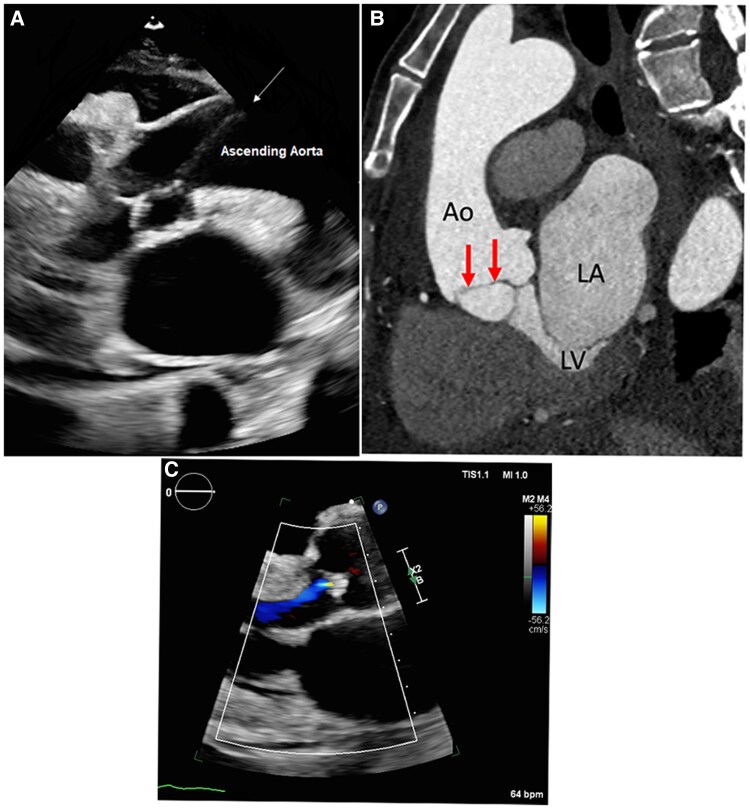 Figure 1
Peer Reviews
No public reviews on file for this paper yet. If you reviewed it on a platform where reviews are public (OpenReview, ICLR, NeurIPS, ICML), you can paste yours below so the community can read it here.
Videos
No videos yet. Explain this paper in a talk, walkthrough, or lecture? Add one.
Taxonomy
TopicsCardiac Valve Diseases and Treatments · Cardiac Structural Anomalies and Repair · Aortic Disease and Treatment Approaches
Case description
A 58-year-old female presented to the emergency room with a malignant gastric outlet obstruction. Her physical exam was significant for a new murmur. A transthoracic echocardiogram (TTE) was completed demonstrating linear echoes within the aorta suggestive of dissection in the aortic arch (Figure 1A). An emergent computed tomography angiogram (CTA) chest was obtained given the suspicion of a Type B aortic dissection but was limited due to motion artefact. A CTA-gated chest was obtained, showing a thin non-calcified linear density in the right coronary sinus seen intermittently during the cardiac cycle likely representing a supra-aortic tendon, mimicking a dissection flap (Figure 1B). Given that this was a benign finding, and the patient wished to pursue hospice, further interventions were not in line with her goals.
(A) Transthoracic echocardiogram with concern for linear echoes within the aorta suggestive of dissection (white arrow). (B) Sagittal oblique reformatted multiplanar image from a retrospectively gated cardiac computed tomography angiogram: There is a thin linear filling defect (inferior to Ao) along the right aortic cusp seen intermittently during the cardiac cycle without any associated aortic wall haematoma or irregularity. This corresponds to the abnormality seen on echo. (C) Mild aortic regurgitation demonstrated on transthoracic echocardiography. This is the most common finding associated with fibrous band. Ao, aorta; LA, left atrium; LV, left ventricle.
Supra-aortic tendons have been described as congenital fibrous or elastic bands typically found during autopsy or incidentally on imaging. An explanation for fibrous band formation is the incomplete remodelling of cardiac cushions during aortic valve development.^1^ They are often associated with other congenital anomalies such as bicuspid and quadricuspid valves and aortic regurgitation with and without band ruptures.^2^ In one study, a fibrous band identified on transoesophageal echocardiography had a sensitivity of 57% and 92% specificity for aortic cusp prolapse, with the most common cusp being right cusp.^3^ Our patient also exhibited mild aortic regurgitation (AR), with the fibrous band appearing to originate from the right coronary sinus (Figure 1C).
Current guidelines do not highlight the presence of bands as a significant concern for AR; however, they may be useful tools for identifying patients with AR and cusp prolapse.^4^ Management of incidental fibrous bands is poorly defined in the literature due to their rarity and asymptomatic nature. Suggested approaches include follow-up with TTE and surgery consultation if significant aortic valve dysfunction is present.
Consent: The authors confirm that written consent was obtained for publication, and all identifiable patient information has been removed from images in compliance with COPE guidelines.
Funding: None declared.
The reference list from the paper itself. Each links out to its DOI / PubMed record.
- 1Ajithdoss DK, Arenas-Gamboa AM, Edwards JF. A fibrous band associated with the non-coronary aortic valve cusp in a dog. J Vet Cardiol 2011;13:127–129.21641896 10.1016/j.jvc.2011.01.004 · doi ↗ · pubmed ↗
- 2Minami H, Asada T, Gan K, Yamada A, Sato M. Aortic regurgitation caused by rupture of the abnormal fibrous band between the aortic valve and aortic wall. Gen Thorac Cardiovasc Surg 2011;59:488–490.21751110 10.1007/s 11748-010-0722-8 · doi ↗ · pubmed ↗
- 3Boodhwani M, de Kerchove L, Watremez C, Glineur D, Vanoverschelde JL, Noirhomme P, et al Assessment and repair of aortic valve cusp prolapse: implications for valve-sparing procedures. J Thorac Cardiovasc Surg 2011;141:917–925.21292284 10.1016/j.jtcvs.2010.12.006 · doi ↗ · pubmed ↗
- 4Otto CM, Nishimura RA, Bonow RO, Carabello BA, Erwin JP, Gentile F, et al 2020 ACC/AHA guideline for the management of patients with valvular heart disease: a report of the American College of Cardiology/American Heart Association joint committee on clinical practice guidelines. Circulation 2021;143:e 72–e 227.33332150 10.1161/CIR.0000000000000923 · doi ↗ · pubmed ↗